. 2012 Jun 15;188(12):6156-64.
doi: 10.4049/jimmunol.1102604. Epub 2012 May 16.

Augmented IL-15Rα expression by CD40 activation is critical in synergistic CD8 T cell-mediated antitumor activity of anti-CD40 antibody with IL-15 in TRAMP-C2 tumors in mice

Affiliations

Augmented IL-15Rα expression by CD40 activation is critical in synergistic CD8 T cell-mediated antitumor activity of anti-CD40 antibody with IL-15 in TRAMP-C2 tumors in mice

Meili Zhang et al. J Immunol. .

Abstract

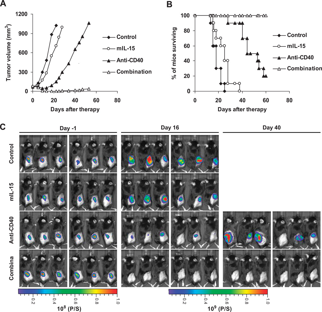
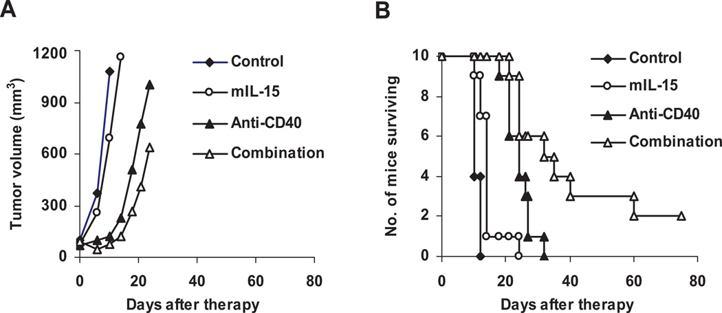
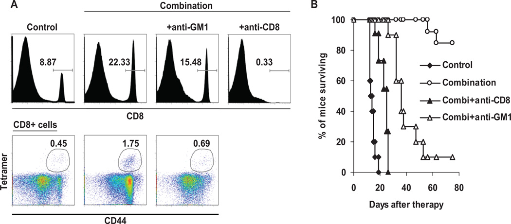
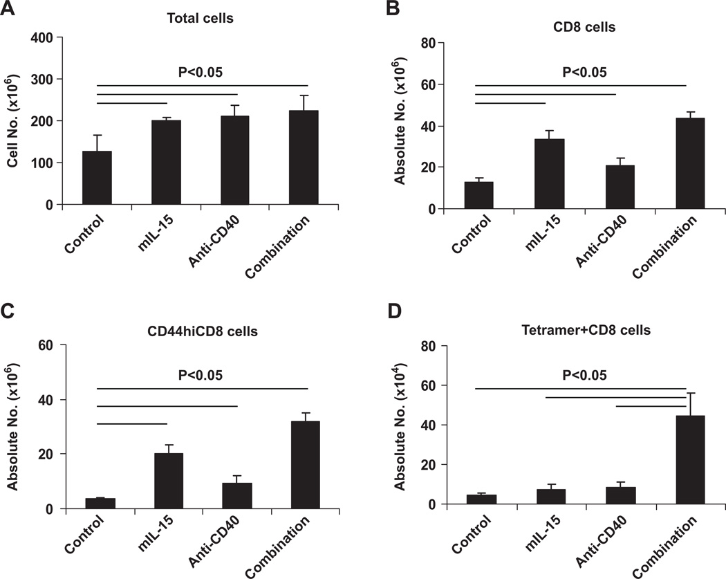
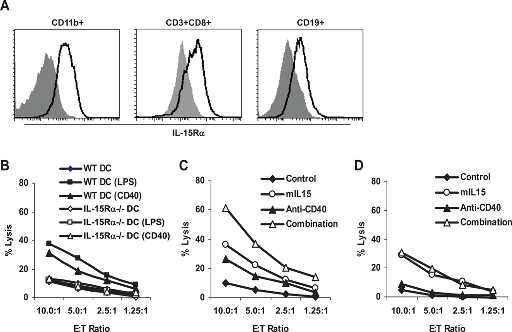
IL-15 has potential as an immunotherapeutic agent for cancer treatment because it is a critical factor for the proliferation and activation of NK and CD8(+) T cells. However, monotherapy of patients with malignancy with IL-15 that has been initiated may not be optimal, because of the limited expression of the private receptor, IL-15Rα. We demonstrated greater CD8 T cell-mediated therapeutic efficacy using a combination regimen of murine IL-15 administered with an agonistic anti-CD40 Ab (FGK4.5) that led to increased IL-15Rα expression on dendritic cells (DCs), as well as other cell types, in a syngeneic established TRAMP-C2 tumor model. Seventy to one hundred percent of TRAMP-C2 tumor-bearing wild-type C57BL/6 mice in the combination group manifested sustained remissions, whereas only 0-30% in the anti-CD40-alone group and none in the murine IL-15-alone group became tumor free (p < 0.001). However, the combination regimen showed less efficacy in TRAMP-C2 tumor-bearing IL-15Rα(-/-) mice than in wild-type mice. The combination regimen significantly increased the numbers of TRAMP-C2 tumor-specific SPAS-1/SNC9-H(8) tetramer(+)CD8(+) T cells, which were associated with the protection from tumor development on rechallenge with TRAMP-C2 tumor cells. Using an in vitro cytolytic assay that involved NK cells primed by wild-type or IL-15Rα(-/-) bone marrow-derived DCs, we demonstrated that the expression of IL-15Rα by DCs appeared to be required for optimal IL-15-induced NK priming and killing. These findings support the view that anti-CD40-mediated augmented IL-15Rα expression was critical in IL-15-associated sustained remissions observed in TRAMP-C2 tumor-bearing mice receiving combination therapy.


Conflict of interest statement

Conflict of interest: No conflict of interest exists.

Figures

Figure 1
Figure 1. The combination therapy that involved mIL-15 and an agonistic anti-CD40 antibody led to the regression of established TRAMP-C2 tumors in wild type C57BL/6 mice
The therapy was started when the tumors were well established with an average volume of 80mm3. (A) Changes of tumor volumes during the therapeutic course and (B) Kaplan-Meier survival plot of the mice in one of the two therapeutic studies (n=9–10). Treatment with mIL-15 alone (○) at a dose of 2.5µg/mouse, 5 days a week for 2 weeks inhibited the tumor growth slightly and prolonged survival of the TRAMP-C2 tumor-bearing mice when compared with the mice in the PBS control group (♦) (p<0.05) while treatment with the anti-CD40 antibody (▲) at a dose of 200 µg/mouse on day 0, then 100 µg/mouse on days 3, 7 and 10 provided greater inhibition of the tumor growth and prolonged survival of the TRAMP-C2 tumor-bearing mice when compared with the mice in the PBS control or mIL-15 alone group (p<0.001). Furthermore, combination therapy (△) with both mIL-15 and the anti-CD40 antibody provided a greater therapeutic efficacy as demonstrated by the fact that all of the mice in the combination group were alive at day 60 with 80% becoming tumor free while only 20% of the mice in the anti-CD40 antibody alone group and none of the mice in the PBS control and mIL-15 alone groups were alive at that time. (C). Bioluminescence imaging of the TRAMP-C2/luc-GFP tumor-bearing mice confirmed efficacy of combination treatment. Groups of 6 mice bearing TRAMP-C2/luc-GFP tumors were treated using the same therapeutic protocol. The bioluminescence images were taken at different time points. Treatment with anti-CD40 antibody significantly delayed the tumor growth. However, at day 40 after initiation of the therapy, only one out of 6 mice in the anti-CD40 group was tumor free. In contrast, the combination treatment led to the regression of the tumors with all of the 6 mice in the group becoming and remaining tumor free.
Figure 2
Figure 2. TRAMP-C2 tumor-bearing IL-15Rα−/− mice showed reduced efficacy with the combination regime when compared to wild type mice
(A) Changes of tumor volumes during the therapeutic course and (B) Kaplan-Meier survival plot of the mice in one of the two therapeutic studies (n=10). The protocol for this study was the same as that in Figure1. Treatment with mIL-15 alone (○) showed very little therapeutic efficacy in the TRAMP-C2 tumor-bearing IL-15Rα −/− mice. Treatment with the anti-CD40 antibody (▲) significantly delayed the tumor growth and prolonged the survival of the TRAMP-C2 tumor-bearing mice when compared with the mice in either PBS control (♦) or mIL-15 alone group (p<0.001). Furthermore, combination therapy (△) with both mIL-15 and the anti-CD40 antibody provided a greater therapeutic efficacy as seen by the prolonged survival with 20% of the mice becoming tumor free whereas none of the mice in other groups was tumor free (p<0.001). The results were from one of two experiments.
Figure 3
Figure 3. CD8+ T-cells played a major role in the combination regimen mediated antitumor activity in the TRAMP-C2 model
TRAMP-C2 tumor-bearing mice were treated with anti-asialo-GM1 (50 µL) or purified rat anti-mouse CD8 antibody (200 µg/injection), together with the combination regimen of mIL-15 and the anti-CD40 antibody. (A) Representative FACS analysis of PBMC from the mice at day 19 after initiation of the therapy. The results showed that the addition of an anti-CD8 antibody was associated with the depletion of more than 90% of CD8+ cells. Administration of anti-asialo-GM1 reduced the number of CD8+ cells, especially CD44highCD8+ and TRAMP-C2 tumor specific SPAS-1/SNC9-H8 tetramer+CD8+ cells. (B) Kaplan-Meier survival plot of the mice. Compared with the group (○) receiving the combination therapy alone, depletion of CD8+ T-cells (▲) with the anti-CD8 antibody in association with combination treatment nearly abrogated the antitumor efficacy (p<0.001) and administration of anti-asialo-GM1 (△) reduced the antitumor activity (p<0.001). The experiment was repeated and the results of the two experiments were pooled together.
Figure 4
Figure 4. The combination of mIL-15 with the anti-CD40 antibody led to a significant increase in the absolute numbers of TRAMP-C2 tumor specific SPAS-1/SNC9-H8 tetramer+CD8+ cells in the spleens of TRAMP-C2 tumor-bearing wild type mice
TRAMP-C2 tumor-bearing mice were treated using the same therapeutic protocol as used in Figure 1. At day 12 after initiation of the therapy, all of the mice (4 mice/group) were sacrificed and spleens were taken. Splenocytes were separated and counted. The percentages of CD8+, CD44highCD8+, SPAS-1/SNC9-H8 tetramer+CD8+ were analyzed by flow cytometry. (A) Total cell numbers per spleen. (B) Absolute numbers of CD8+ cells per spleen. (C) Absolute numbers of CD44highCD8+ cells per spleen. (D) Absolute numbers of SPAS-1/SNC9-H8 tetramer+CD8+ cells per spleen. Treatment with mIL-15 or anti-CD40 antibody or their combination increased the total cell numbers and the absolute numbers of CD8+ cells and CD44highCD8+ cells in the spleen when compared with the control group (p<0.05). Critically, only combination regimen significantly increased the numbers of SPAS-1/SNC9-H8 tetramer+CD8+ cells when compared with all other groups (p<0.05).
Figure 5
Figure 5. Enhanced TRAMP-C2 tumor specific SPAS-1/SNC9-H8 tetramer+CD8+ T-cells were associated with protection from a rechallenge with TRAMP-C2 tumor cells
(A) Kaplan-Meier survival plot of mice that received rechallenge of TRAMP-C2 cells at day 40 after initiation of the original therapy. When compared with control mice (♦) which did not receive TRAMP-C2 cells and any treatment previously, mice surviving the first challenge with TRAMP-C2 cells (○) demonstrated resistance to TRAMP-C2 tumor development (p<0.001). Depletion of CD8 cells (▲) nearly abrogated protection mediated by combination therapy (p<0.01). (B) Kaplan-Meier survival plot of mice that received rechallenge of MC38 cells at day 40 after original therapy. Mice surviving the challenge with TRAMP-C2 cells did not show resistance to an irrelevant tumor, MC38, development (p>0.5). (C) Representative FACS analysis of PBMC from mice just before rechallenge. Mice that initially received the TRAMP-C2 tumor that became and remained tumor free after receiving the combination regimen manifested an increase in the percentages of CD8+, CD44highCD8+ T cells, especially the SPAS-1/SNC9-H8 tetramer+CD8+ T-cells when compared with mice in the control group that had not received either the TRAMP-C2 tumor cells or any treatment previously. (D) Bioluminescence images. Surviving tumor free mice in the combination group from Fig. 1C were rechallenged with TRAMP-C2/luc-GFP cells at day 40 after initiation of the therapy. The mice surviving the first challenge with TRAMP-C2/luc-GFP cells demonstrated resistance to the tumor development when compared with the mice in the control group.
Figure 6
Figure 6. CD40 activation induced IL-15Rα expression and increased levels of IL-15Rα on the BMDCs appear critical for NK cell lytic activity
(A) Histograms of flow cytometry analysis of IL-15Rα expression on cell surfaces. C57BL/6 mice were treated with PBS or with the anti-CD40 antibody at a dose of 200µg on day 0, followed by 100µg on days 3. At day 5, the splenocytes were separated from the mice for flow cytometry analysis. Treatment with the anti-CD40 antibody increased the expression of IL-15Rα (solid line) on all cell types tested (CD19+, CD11b+ and CD3+CD8+ cells) when compared with that observed in the mice treated with PBS (grey shade). (B) Cytolytic activities of NK cells primed by wild type or IL-15Rα−/− BMDCs stimulated by LPS, anti-CD40 or medium alone. Compared with BMDCs cultured with medium, wild type BMDCs stimulated with LPS or anti-CD40 antibody enhanced cytolytic activity of NK cells while IL-15Rα−/− comparably stimulated BMDCs did not. (C) Cytolytic activities of NK cells cocultured with wild type BMDCs in presence or absence of anti-CD40 antibody or mIL-15 or their combination. With anti-CD40 antibody or mIL-15, NK cells showed enhanced cytolytic activity toward target cells when compared with NK cells cocultured with BMDCs in medium alone. Furthermore, with both anti-CD40 antibody and mIL-15, NK cells showed greatest cytolytic activity toward target cells when compared with NK cells in all other cocultures. (D) Cytolytic activities of NK cells cocultured with IL-15Rα−/− BMDCs in presence or absence of anti-CD40 antibody or mIL-15 or their combination. In contrast to wild type BMDCs, NK cells cocultured with IL-15Rα−/− BMDCs in presence of anti-CD40 antibody did not show increased lysis activity toward target cells when compared with NK cells cocultured with IL-15Rα−/− BMDCs in medium alone. With both anti-CD40 antibody and mIL-15, NK cells only showed a comparably increased lysis activity toward target cells as NK cells cocultured with IL-15Rα−/− BMDCs in presence of mIL-15 alone, suggesting that expression of IL-15Rα by BMDCs is critical for the synergetic effect of the combination regimen on activation of NK cells.

Similar articles

Cited by

References

    1. Heemskerk B, Liu K, Dudley ME, Johnson LA, Kaiser A, Downey S, Zheng Z, Shelton TE, Matsuda K, Robbins PF, Morgan RA, Rosenberg SA. Adoptive cell therapy for patients with melanoma, using tumor-infiltrating lymphocytes genetically engineered to secrete interleukin-2. Hum. Gene. Ther. 2008;19:496–510. - PMC - PubMed
    1. Klapper JA, Downey SG, Smith FO, Yang JC, Hughes MS, Kammula US, Sherry RM, Royal RE, Steinberg SM, Rosenberg S. High-dose interleukin-2 for the treatment of metastatic renal cell carcinoma : a retrospective analysis of response and survival in patients treated in the surgery branch at the National Cancer Institute between 1986 and 2006. Cancer. 2008;113:293–301. - PMC - PubMed
    1. Diab A, Cohen AD, Alpdogan O, Perales MA. IL-15: targeting CD8+ T cells for immunotherapy. Cytotherapy. 2005;7:23–35. - PubMed
    1. Evans R, Fuller JA, Christianson G, Krupke DM, Troutt AB. IL-15 mediates anti-tumor effects after cyclophosphamide injection of tumor-bearing mice and enhances adoptive immunotherapy: the potential role of NK cell subpopulations. Cell Immunol. 1997;179:66–73. - PubMed
    1. Kobayashi H, Dubois S, Sato N, Sabzevari H, Sakai Y, Waldmann TA, Tagaya Y. Role of trans-cellular IL-15 presentation in the activation of NK cell-mediated killing, which leads to enhanced tumor immunosurveillance. Blood. 2005;105:721–727. - PubMed

Publication types

MeSH terms