. 2011 Sep;2(9):900-9.
doi: 10.1177/1947601911431885.

Transcriptional Regulation of EGR1 by EGF and the ERK Signaling Pathway in Prostate Cancer Cells

Affiliations

Transcriptional Regulation of EGR1 by EGF and the ERK Signaling Pathway in Prostate Cancer Cells

Jennifer Gregg et al. Genes Cancer. 2011 Sep.

Abstract

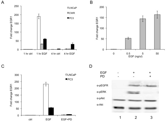
The early growth response gene 1, EGR1, is an important transcriptional regulator and acts as the convergent point between a variety of extracellular stimuli and activation of target genes. Unlike other tumor types, prostate tumors express high levels of EGR1 relative to normal tissues. However, the mechanism of EGR1 regulation in prostate tumor cells is unknown. As EGR1 expression and epidermal growth factor (EGF) signaling are frequently upregulated in prostate tumors, we tested the hypothesis that EGF induces EGR1 expression in prostate cancer cells. Using RT-PCR to quantify EGR1 transcripts, we found that EGF induced EGR1 expression in a dose- and time-dependent manner and the ERK pathway inhibitor, PD98059, abrogated the EGF-mediated EGR1 response in LNCaP and PC3 cells. Analysis of the EGR1 promoter using deletion constructs identified an EGF-responsive region in the proximal promoter (-771 to -245 bp) containing 3 potential serum response element (SRE) sites. In vivo chromatin immunoprecipitation assays demonstrated that Elk-1 binding at the SRE sites of the EGR1 promoter was enhanced by EGF treatment in PC3 cells. Overexpression of Elk-1 was sufficient to activate the EGF-responsive region of EGR1 promoter in PC3 cells and, similarly, a dominant-negative Elk-1 suppressed EGR1 promoter activity. Taken together, these results demonstrate for the first time that EGR1 expression in PC3 cells is mediated through an EGF-ERK-Elk-1 signaling cascade.

Keywords: EGF; EGR1; Elk-1; prostate cancer.


Conflict of interest statement

The author(s) declared no potential conflicts of interest with respect to the research, authorship, and/or publication of this article.

Figures

Figure 1.
Figure 1.
EGF elevates EGR1 transcript levels via the ERK pathway in prostate cancer cells. (A) EGF activates EGR1 transcript levels in a time and dose dependent manner. LNCaP, CWR, and PC3 cells were serum deprived overnight and treated with 50 ng/mL EGF for the times indicated. RNA was isolated and assayed using RT-PCR to quantify EGR1 transcript levels. Values were normalized to 18S transcript levels and each experiment was done in triplicate. Data are represented as mean ± SEM (n = 3) fold change values relative to 1 h control for each cell type. (B) LNCaP cells were serum deprived overnight and treated with the indicated concentrations of EGF for 1 h. RNA was isolated and assayed as described above. Data are represented as mean ± SEM (n = 3) fold change values relative to 0 ng/mL EGF. (C) EGR1 transcriptional regulation is mediated by the ERK pathway in LNCaP and PC3 cells. Cells were pretreated with 50 µM PD98059 for 1 h followed by treatment with 50 ng/mL EGF for an additional h. EGR1 transcript levels were measured as described above. Data are represented as mean ± SEM (n = 3) fold change values relative to control for each cell type. (D) Phosphorylation of ERK, Akt, and EGFR in LNCaP cells. LNCaP cells were serum-deprived overnight and pretreated with 50 µM PD98059 for 1 h followed by treatment with 50 ng/mL EGF or vehicle (PBS) for 15 min. Protein lysates were separated by SDS-PAGE and probed with phosphospecific antibodies using standard Western blot techniques. Signal was detected with luminol reagent and a FujiFilm LAS-3000 detection system. Akt antibody was used as a loading control.
Figure 2.
Figure 2.
Transcriptional activation of the hEGR1 promoter is dependent on the proximal SRE sites and the Erk pathway in LNCaP and PC3 cells. (A) Schematic representation of the EGR1-luc promoter deletion constructs. Predicted Elk-1 and SRF transcription factor binding sites were identified using MatInspector (Genomatix, Munich, Germany) software. Clusters of core (c), proximal (p), and distal (d) SRF/Elk-1 sites are indicated. (B) Transcriptional activation of the hEGR1 promoter is dependent on the −771 to −245 region containing the proximal SRE sites. Cells were transfected with 500 ng of the appropriate luciferase construct, grown for 24 h, serum deprived overnight, and then treated with either vehicle control or 5 ng/mL EGF (LNCaP) or 50 ng/mL (PC3) for 5 h. (C and D) LNCaP and PC3 cells, respectively, were cultured as described above but pretreated with or without 50 µM PD98059 for 1 h prior to transfection with 500 ng of the appropriate luciferase construct. Luciferase activity was measured and normalized to protein concentration. Data are reported as fold activation relative to vehicle treated −1,260 hEGR1 control. The data represents the average fold activation of relative light units ± SEM of 3 experiments repeated at least twice with similar results.
Figure 3.
Figure 3.
Chromatin immunoprecipitation analysis of the proximal region of SRE sites within the EGR1 promoter in PC3 cells. (A) Arrows depict primers designed to flank the proximal region of SRE sites (–603 to −315 bp) within the EGR1 promoter. SRF/Elk-1 sites are depicted as in Figure 3A. (B) Chromatin obtained from PC3 (top panel) and LNCaP (bottom panel) cells treated as described in Figure 1 was immunoprecipitated and amplified by PCR as described in the text. Lane 1, DNA marker. Immunoprecipitations in the absence (–) or presence (+) of EGF using 1 µg of negative control IgG (lanes 2 and 3) or anti-Elk-1 Ab (lanes 4 and 5). Lanes 6 and 7 represent PCR amplification of 1% input chromatin (nonimmunoprecipitated). (C) Quantitation of Elk-1 binding at the proximal promoter of EGR1 in PC3 cells by RT-PCR. Immunoprecipitated and control input DNA was quantified using SYBR-green master mix and the same primers as described in (A). Data were normalized relative to nonimmunoprecipitated input using adjusted Ct values and is represented as percentage of input DNA.
Figure 4.
Figure 4.
Activation of the −771 hEGR1 promoter is enhanced by Elk-1 expression and inhibited by a dominant negative Elk-1 in PC3 cells. Cells were grown and treated with EGF as previously described in Figure 2 above. (A) Cells were co-transfected with 250 ng of the −771 hEGR1-luc promoter construct and either 500 ng of full-length Elk-1, pcDNA3 empty vector, or Elk-1ΔDBD, containing a deletion of the Elk-1 DNA-binding domain. (B) PC3 cells were co-transfected with 250 ng of the −771 hEGR1-luc promoter construct and either 500 ng of pcDNA3 empty vector or the indicated concentrations of the Elk-En expression construct. Data are reported as fold activation relative to pcDNA3 control. Luciferase activity is represented as the average fold activation of relative light units ± SEM of 3 experiments repeated at least twice with similar results.

References

    1. Yan SF, Fujita T, Lu J, et al. Egr-1, a master switch coordinating upregulation of divergent gene families underlying ischemic stress. Nat Med. 2000;6:1355-61 - PubMed
    1. Thiel G, Cibelli G. Regulation of life and death by the zinc finger transcription factor egr-1. J Cell Physiol. 2002;193:287-92 - PubMed
    1. Adamson ED, Mercola D. Egr1 transcription factor: Multiple roles in prostate tumor cell growth and survival. Tumor Biol. 2002;23:93-102 - PubMed
    1. Levin WJ, Press MF, Gaynor RB, et al. Expression patterns of immediate early transcription factors in human non-small cell lung cancer. Oncogene. 1995;11:1261-9 - PubMed
    1. Joslin JM, Fernald AA, Tennant TR, et al. Haploinsufficiency of EGR1, a candidate gene in the del(5q), leads to the development of myeloid disorders. Blood. 2007;110:719-26 - PMC - PubMed

LinkOut - more resources