. 2012 Sep;12(9):2322-34.
doi: 10.1111/j.1600-6143.2012.04090.x. Epub 2012 May 17.

Lymphotoxin-beta receptor blockade induces inflammation and fibrosis in tolerized cardiac allografts

Affiliations

Lymphotoxin-beta receptor blockade induces inflammation and fibrosis in tolerized cardiac allografts

Y Nakayama et al. Am J Transplant. 2012 Sep.

Abstract

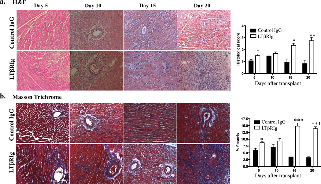
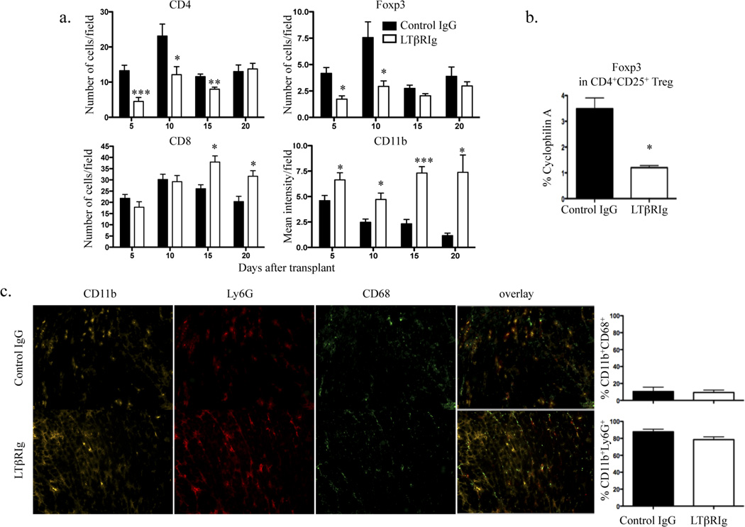
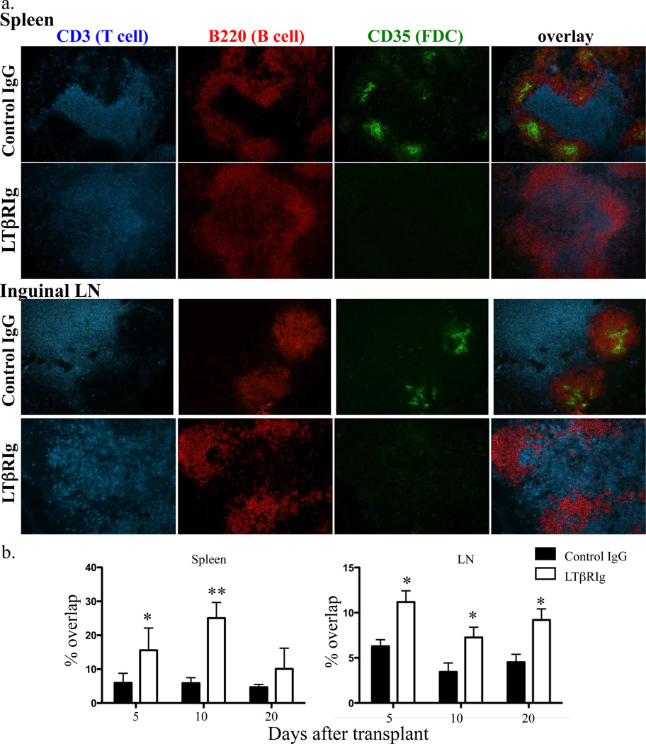
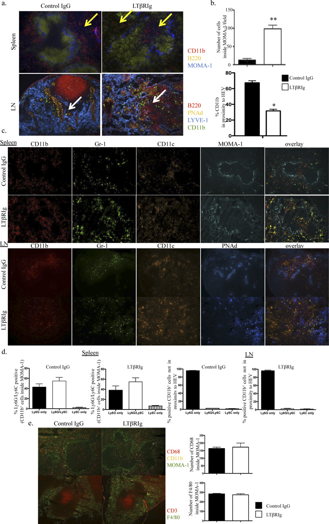
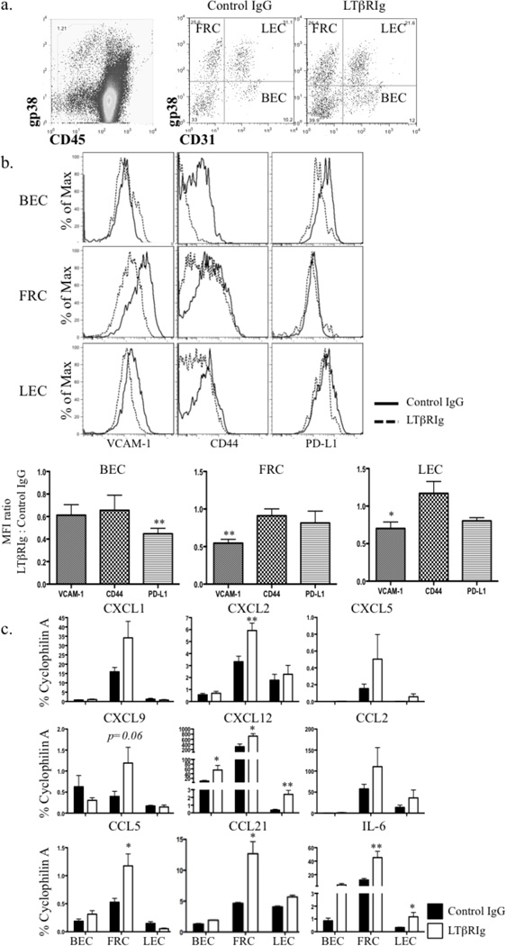
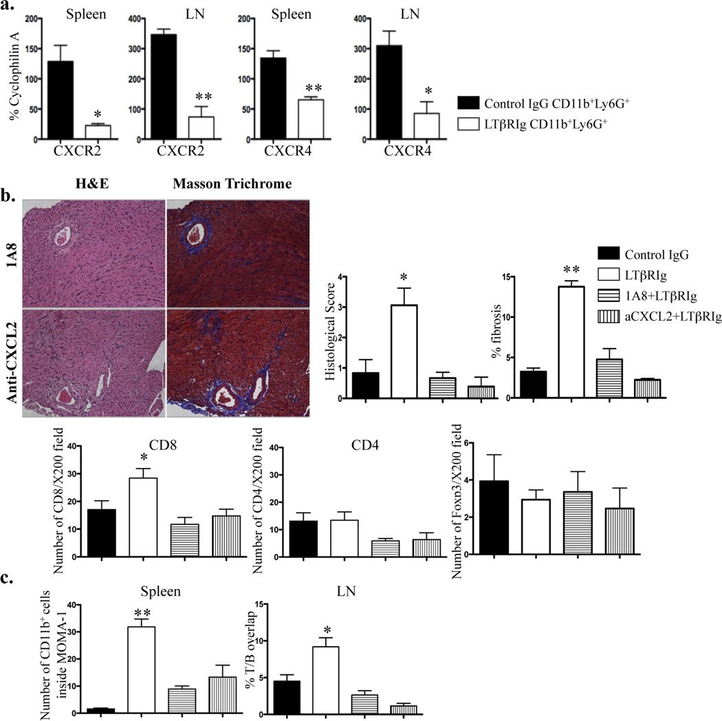
The lymphotoxin system (LT) regulates interactions between lymphocytes and stromal cells to maintain lymphoid microenvironmental homeostasis. Soluble LT beta-receptor-Ig (LTβRIg) blocks lymphocyte LTα1β2-stromal cell LTβR signaling. In a murine cardiac allograft model, LTbRIg treatment reversed the tolerance induced by anti-CD40L antibody leading to graft inflammation and fibrosis. LTβRIg treatment decreased PD-L1 expression by blood endothelial cells, and decreased VCAM-1 while increasing CXCL1, CXCL2, CXCL12, CCL5, CCL21 and IL-6 expression in fibroblastic reticular cells. In secondary lymphoid organs these effects caused T- and B cell zone disruption, loss of CD35(+) follicular dendritic cells and abnormal recruitment of CD11b(+) Ly6G(+) neutrophils. These disruptions correlated with increased numbers of CD8(+) T cells and CD11b(+) Ly6G(+) neutrophils, and decreased numbers of CD4(+) T cells and Foxp3(+) regulatory T cells in the grafts. Depleting neutrophils or blocking neutrophil-attracting chemokines restored normal histology in lymph node, spleen and grafts. Taken together, LTβRIg treatment altered stromal subset, particularly fibroblastic reticular cell, production of cytokines and chemokines, resulting in changes in neutrophil recruitment in spleen, lymph node and grafts, and inflammation and fibrosis associated with decreased Foxp3(+) regulatory T cells and increased CD8(+) T cell infiltration of grafts.


Figures

Figure 1
Figure 1
LTβRIg causes chronic inflammation and fibrosis in grafts. Grafts from tolerized C57BL/6 recipients treated with control IgG or LTβRIg were harvested on the indicated days and stained with H&E (a) or Masson Trichrome stain (b). Representative images are shown. Magnification X200. Histological scores (a) and percent fibrosis (b) for indicated days were measured. Results from 4–6 mice/group, 2–8 sections/graft, and 3–10 fields/section. * p<0.05, ** p<0.001, *** p<0.0001.
Figure 2
Figure 2
Increased numbers of CD8+ T cells and CD11b+ graft infiltrating cells accompany LTβRIg treatment. a. Numbers of CD8+, CD4+, and Foxp3+ T cells and the mean intensity of CD11b+ cells in the graft sections on the indicated days determined by Image J software. Results from 4–6 mice/group, 4–6 sections/graft, and 4 to 27 fields/section. b. CD4+CD25+ Treg were sorted on day 5 from grafts and RNA was isolated for qRT-PCR. Results are from 3 sortings, 3–4 mice/group/experiment. c. Frozen sections of grafts stained for CD11b, Ly6G, and CD68. Representative images from day 10. Magnification X200. Quantitative analysis of images for percentages of CD11b+CD68+ and CD11b+Ly6G+ cells. 2–4 section/graft, 4–5 mice/group. * p<0.05, ** p<0.005, *** p<0.0001.
Figure 3
Figure 3
LTβRIg causes structural changes in spleen and LN. a. Spleen and LN were harvested 5, 10, and 20 days after transplant, and stained for CD3, B220, and CD35. Representative images from day 5 samples. Magnification for spleen X100, LN X200. 3–4 mice/group. b. Percentages of T and B cell zone overlap in the images in (a) were measured by Volocity software. Results are from 3–4 mice/group; 2 sections/spleen with 4–8 fields/spleen section; and 2 LN/mouse, 2 sections/LN with 3–6 fields/LN section. * p< 0.05, ** p<0.005.
Figure 4
Figure 4
LTβRIg alters CD11b+ cell distribution. a. Spleen (top) and LN (bottom) were harvested 10 days after transplant. Spleens were stained for CD11b (red), B220 (yellow), and MOMA-1 (blue). Yellow arrows indicate CD11b+ cells. LN were stained for B220 (red), PNAd (HEV) (yellow), LYVE-1 (blue), and CD11b (green). White arrows indicate CD11b+ cells. Magnification, spleen X100, LN X200. b. Numbers of CD11b+ cells inside WP in spleen were measured with Volocity software (top). Percentages of CD11b+ cells in close proximity to the HEV in LN were quantitated with Volocity software (bottom). c. Spleens from day 10 stained for CD11b (red), Gr-1 (blue), CD11c (orange) and MOMA-1 (green). LN were stained for CD11b (red), Gr-1 (green), CD11c (orange), and PNAd (blue). Magnification, spleen X100, LN X200. d. Percentages of Ly6G and Ly6C positive cells in the populations measured in spleen (top) and LN (bottom). Results are from 4–6 mice/group; 2 spleen sections/mouse, 4–8 fields/spleen section; and 2 LNs/mouse, 2 sections/LN, 3–8 fields/LN section. e. Spleen sections from day 5 stained for CD11b, CD68, CD3, and F4/80. The number of CD68+ cells inside WP and the number of F4/80+ cells per field were determined by Volocity software. Magnification X100. * p<0.005 ** p<0.0001.
Figure 5
Figure 5
LTβRIg treatment alters LNSC phenotype. a. Representative dot plots of LNSC. LNSC were CD45 (left), and separated into 4 subsets based on CD31 and gp38 expression (right). b. FRC, LEC, and BEC gated on CD31 and gp38; and PD-L1, CD44 and VCAM-1 assayed by flow cytometry on day 5, 10 and 30 (day 10 shown). Histograms (top) and mean intensity (bottom) for each marker. Results are normalized to naive and IgG controls. Results representative from three experiments with 1–6 mice/group. c. mRNA from each subset was isolated on day 10 after transplantation and qRT-PCR performed for the indicated genes. Results from 4 experiments with LN from 3–4 mice pooled/experiment. * p<0.05 ** p<0.005.
Figure 6
Figure 6
Neutrophil presence is associated with graft inflammation and fibrosis. a. Neutrophils were sorted from spleen and LN on day 5 after transplant. qRT-PCR was performed for indicated molecules. Results from 2 mice/group, 2 independent experiments. b. Grafts from tolerogen treated C57BL/6 recipients treated with control IgG or LTβRIg were injected with either 1A8 (top) or anti-CXCL2 (bottom) antibody, harvested on 20 and stained with H&E (left) or Masson Trichrome stain (right). Histological score and percent fibrosis, and numbers of CD8+, CD4+ and Foxp3+ cells on day 20 for control IgG and LTβRIg treated grafts are from Figures 1 and 2. Representative images are shown. Magnification X200. Results from 3 mice/group, 6 sections/graft, and 4–14 fields/section. c. The number of CD11b+ cells inside WP in spleen, and the percent of T and B cell area overlap in LN are shown, with control IgG and LTβRIg on day 20 from Figures 3 and 4. Results from 3 mice/group, 6 sections/spleen, 3–6 fields/section, 2 sections/LN, 6–8 LN/mouse and 2–9 fields/section. * p<0.05 ** p<0.005.

References

    1. Bashuda H, Shimizu A, Uchiyama M, Okumura K. Prolongation of renal allograft survival by anergic cells: advantages and limitations. Clin Transplant. 2010;24(Suppl 22):6–10. - PubMed
    1. Gorantla VS, Schneeberger S, Brandacher G, Sucher R, Zhang D, Lee WP, et al. T regulatory cells and transplantation tolerance. Transplant Rev (Orlando) 2010;24(3):147–159. - PMC - PubMed
    1. Watson D, Hu M, Zhang GY, Wang YM, Alexander SI. Tolerance induction by removal of alloreactive T cells: in-vivo and pruning strategies. Curr Opin Organ Transplant. 2009;14(4):357–363. - PubMed
    1. Gudmundsdottir H, Turka LA. T cell costimulatory blockade: new therapies for transplant rejection. J Am Soc Nephrol. 1999;10(6):1356–1365. - PubMed
    1. Wekerle T, Kurtz J, Bigenzahn S, Takeuchi Y, Sykes M. Mechanisms of transplant tolerance induction using costimulatory blockade. Curr Opin Immunol. 2002;14(5):592–600. - PubMed

Publication types