

Identification and characterization of the zebrafish pharyngeal arch-specific enhancer for the basic helix-loop-helix transcription factor Hand2
- PMID: 22595513
- PMCID: PMC3676283
- DOI: 10.1016/j.ydbio.2012.05.003
Identification and characterization of the zebrafish pharyngeal arch-specific enhancer for the basic helix-loop-helix transcription factor Hand2
Abstract
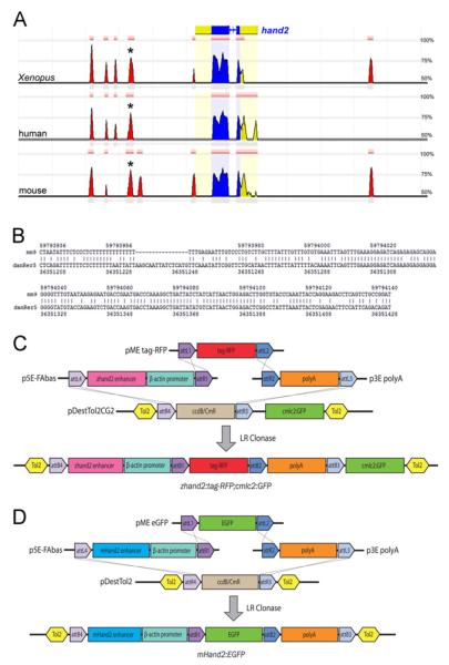
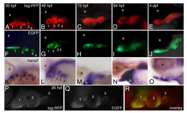
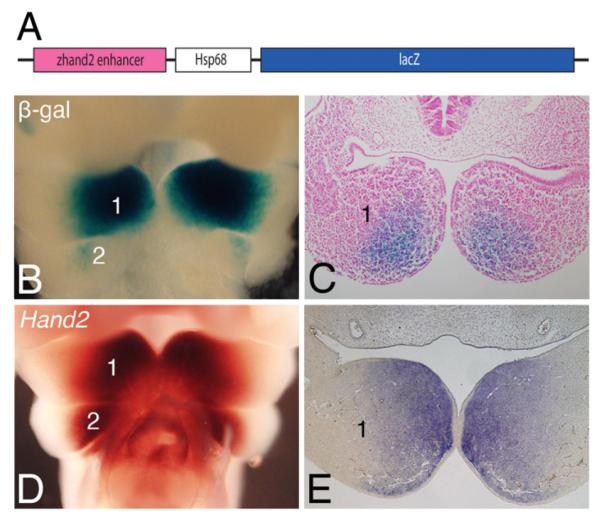
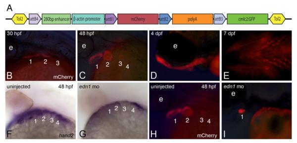
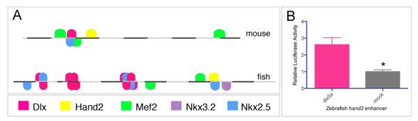
The development of the vertebrate jaw relies on a network of transcription factors that patterns the dorsal-ventral axis of the pharyngeal arches. Recent findings in both mouse and zebrafish illustrate that the basic-helix-loop-helix transcription factor, Hand2, is crucial in this patterning process. While Hand2 has functionally similar roles in these two species, little is known about the regulatory sequences controlling hand2 expression in zebrafish. Using bioinformatics and Tol2-mediated transgenesis, we have generated zebrafish transgenic reporter lines in which either the mouse or zebrafish arch-specific hand2 enhancer direct expression of a fluorescent reporter. We find that both the mouse and zebrafish enhancers drive early reporter expression in a hand2-specific pattern in the ventral pharyngeal arches of zebrafish embryos. These lines provide useful tools to follow ventral arch cells during vertebrate jaw development while also allowing dissection of hand2 transcriptional regulation during this process.
Copyright © 2012 Elsevier Inc. All rights reserved.
Figures
Similar articles
-
Two endothelin 1 effectors, hand2 and bapx1, pattern ventral pharyngeal cartilage and the jaw joint.Development. 2003 Apr;130(7):1353-65. doi: 10.1242/dev.00339. Development. 2003. PMID: 12588851
-
edn1 and hand2 Interact in early regulation of pharyngeal arch outgrowth during zebrafish development.PLoS One. 2013 Jun 24;8(6):e67522. doi: 10.1371/journal.pone.0067522. Print 2013. PLoS One. 2013. PMID: 23826316 Free PMC article.
-
hand2 and Dlx genes specify dorsal, intermediate and ventral domains within zebrafish pharyngeal arches.Development. 2010 Aug 1;137(15):2507-17. doi: 10.1242/dev.049700. Epub 2010 Jun 23. Development. 2010. PMID: 20573696 Free PMC article.
-
Understanding endothelin-1 function during craniofacial development in the mouse and zebrafish.Birth Defects Res C Embryo Today. 2004 Jun;72(2):190-9. doi: 10.1002/bdrc.20007. Birth Defects Res C Embryo Today. 2004. PMID: 15269892 Review.
-
A HANDful of questions: the molecular biology of the heart and neural crest derivatives (HAND)-subclass of basic helix-loop-helix transcription factors.Gene. 2003 Jul 17;312:27-40. doi: 10.1016/s0378-1119(03)00669-3. Gene. 2003. PMID: 12909338 Review.
Cited by
-
Dlx5 and Dlx6 can antagonize cell division at the G1/S checkpoint.BMC Mol Cell Biol. 2019 Apr 11;20(1):8. doi: 10.1186/s12860-019-0191-6. BMC Mol Cell Biol. 2019. PMID: 31041891 Free PMC article.
-
Lateral thinking in syndromic congenital cardiovascular disease.Dis Model Mech. 2023 May 1;16(5):dmm049735. doi: 10.1242/dmm.049735. Epub 2023 May 1. Dis Model Mech. 2023. PMID: 37125615 Free PMC article.
-
Development of enhancer-trapping and -detection vectors mediated by the Tol2 transposon in zebrafish.PeerJ. 2019 Apr 30;7:e6862. doi: 10.7717/peerj.6862. eCollection 2019. PeerJ. 2019. PMID: 31106068 Free PMC article.
-
Heart Enhancers: Development and Disease Control at a Distance.Front Genet. 2021 Mar 10;12:642975. doi: 10.3389/fgene.2021.642975. eCollection 2021. Front Genet. 2021. PMID: 33777110 Free PMC article. Review.
-
Evolution in an extreme environment: developmental biases and phenotypic integration in the adaptive radiation of antarctic notothenioids.BMC Evol Biol. 2016 Jun 29;16(1):142. doi: 10.1186/s12862-016-0704-2. BMC Evol Biol. 2016. PMID: 27356756 Free PMC article.
References
-
- Bronner-Fraser M. Origins and developmental potential of the neural crest. Exp. Cell. Res. 1995;218:405–417. - PubMed
-
- Chai Y, Jiang X, Ito Y, Bringas P, Jr., Han J, Rowitch DH, Soriano P, McMahon AP, Sucov HM. Fate of the mammalian cranial neural crest during tooth and mandibular morphogenesis. Development. 2000;127:1671–1679. - PubMed
Publication types
MeSH terms
Substances
Grants and funding
LinkOut - more resources
Full Text Sources
Molecular Biology Databases
