Review
. 2013 Jan;20(1):3-11.
doi: 10.1038/cdd.2012.63. Epub 2012 May 18.

Regulation of lipid stores and metabolism by lipophagy

Affiliations
Review

Regulation of lipid stores and metabolism by lipophagy

K Liu et al. Cell Death Differ. 2013 Jan.

Abstract

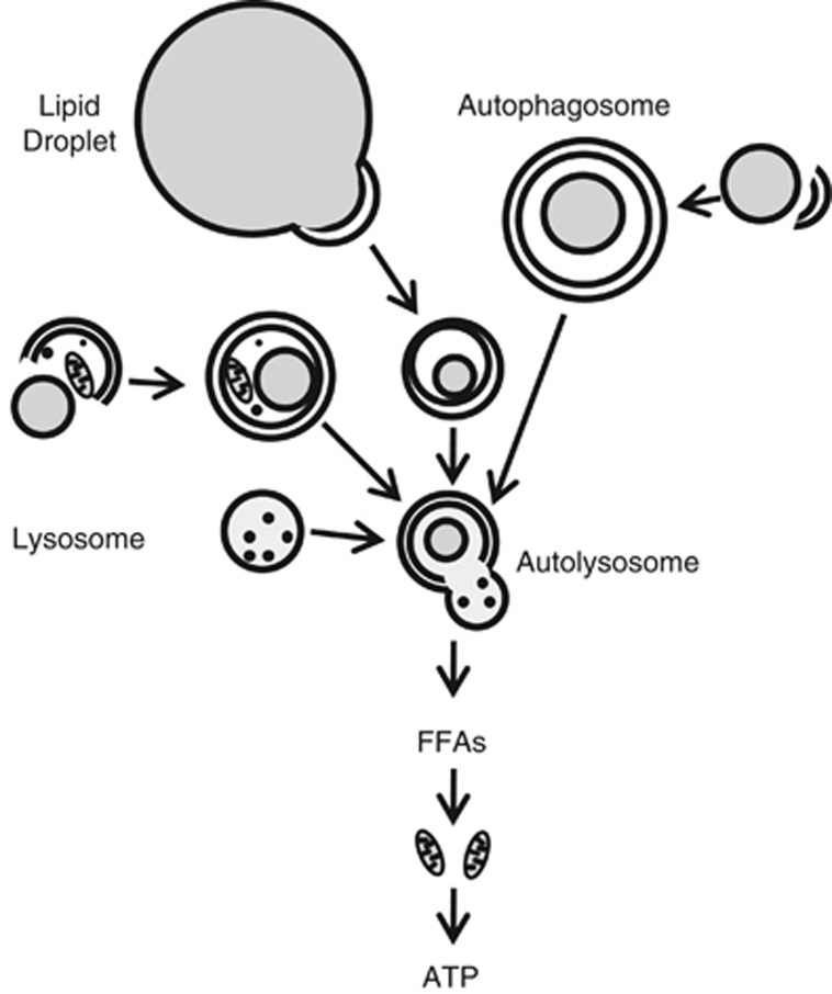
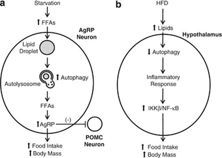
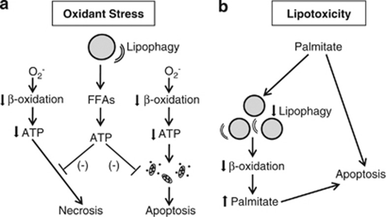
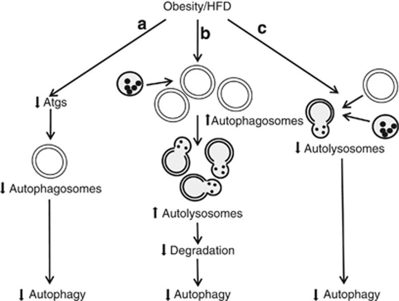
Intracellular lipids are stored in lipid droplets (LDs) and metabolized by cytoplasmic neutral hydrolases to supply lipids for cell use. Recently, an alternative pathway of lipid metabolism through the lysosomal degradative pathway of autophagy has been described and termed lipophagy. In this form of lipid metabolism, LD triglycerides (TGs) and cholesterol are taken up by autophagosomes and delivered to lysosomes for degradation by acidic hydrolases. Free fatty acids generated by lipophagy from the breakdown of TGs fuel cellular rates of mitochondrial β-oxidation. Lipophagy therefore functions to regulate intracellular lipid stores, cellular levels of free lipids such as fatty acids and energy homeostasis. The amount of lipid metabolized by lipophagy varies in response to the extracellular supply of nutrients. The ability of the cell to alter the amount of lipid targeted for autophagic degradation depending on nutritional status demonstrates that this process is selective. Intracellular lipids themselves regulate levels of autophagy by unclear mechanisms. Impaired lipophagy can lead to excessive tissue lipid accumulation such as hepatic steatosis, alter hypothalamic neuropeptide release to affect body mass, block cellular transdifferentiation and sensitize cells to death stimuli. Future studies will likely identify additional mechanisms by which lipophagy regulates cellular physiology, making this pathway a potential therapeutic target in a variety of diseases.


Figures

Figure 1
Figure 1
Process of LD breakdown by lipophagy. Portions of large LDs or entire small LDs are sequestered by a double-membrane autophagosome. Lipids may be sequestered in autophagosomes in combination with other cellular constituents or as the only cargo. Autophagosomes fuse with lysosomes to form autolysosomes in which the substrates of the autophagosome and the hydrolytic enzymes of the lysosome are mixed for cargo degradation. Lipid breakdown leads to release into the cytoplasm of degradation products such as FFAs. FFAs serve to sustain rates of mitochondrial β-oxidation for the generation of ATP to maintain cellular energy homeostasis
Figure 2
Figure 2
Opposing views of the effects of autophagy on hypothalamic food sensing. (a) With acute starvation, the increased serum FFAs released from adipocyte lipid stores result in increased FFA levels in AgRP neurons and FFA incorporation into LDs. Starvation-stimulated autophagy breaks down LDs into FFAs that induce AgRP production. AgRP increases food intake both directly and indirectly through inhibitory effects on POMC neurons. An increase in autophagy in hypothalamic AgRP neurons therefore translates into increased food intake and body mass. (b) With chronic HFD feeding, the hypothalamic levels of autophagy are decreased. Reduced autophagy leads to an inflammatory response, which includes activation of the IκB kinase β (IKKβ)/NF-κB signaling pathway, that increases food intake. In this proposed model, a decrease in hypothalamic neuron autophagy increases food intake and body mass
Figure 3
Figure 3
Two mechanisms for the alteration of cell death responses by lipophagy. (a) Superoxide-induced oxidant stress decreases rates of mitochondrial β-oxidation, leading to decreased cellular ATP content. A profound decrease in ATP leads to cell death from necrosis. A more modest decrease in ATP promotes mitochondrial dysfunction and activation of the mitochondrial death pathway with cytochrome-c release that triggers cell death from apoptosis. Lipophagy mediates resistance to both of these forms of cell death by breaking down LDs into FFAs that can be utilized to make ATP. (b) Lipotoxicity from the saturated FFA palmitate may be mediated in part by its ability to inhibit lipophagy, resulting in decreased levels of β-oxidation and palmitate metabolism, which increase cellular levels of palmitate to trigger apoptosis
Figure 4
Figure 4
Potential mechanisms implicated in the decreased hepatic autophagy that occurs with obesity or HFD feeding. (a) Obesity or HFD may reduce levels of various autophagy factors (Atgs), causing decreased autophagosome formation and levels of autophagy. (b) The liver may form more autophagosomes and autolysosomes, but levels of autophagy decrease nonetheless due to lysosomal defects that preclude degradation. (c) Impaired fusion of autophagosomes and lysosomes may also decrease autophagy

References

    1. Singh R, Kaushik S, Wang Y, Xiang Y, Novak I, Komatsu M, et al. Autophagy regulates lipid metabolism. Nature. 2009;458:1131–1135. - PMC - PubMed
    1. Martin S, Parton RG. Lipid droplets: a unified view of a dynamic organelle. Nat Rev Mol Cell Biol. 2006;7:373–378. - PubMed
    1. Thiele C, Spandl J. Cell biology of lipid droplets. Curr Opin Cell Biol. 2008;20:378–385. - PubMed
    1. Zechner R, Madeo F. Cell biology: another way to get rid of fat. Nature. 2009;458:1118–1119. - PubMed
    1. Marchesini G, Brizi M, Bianchi G, Tomassetti S, Bugianesi E, Lenzi M, et al. Nonalcoholic fatty liver disease: a feature of the metabolic syndrome. Diabetes. 2001;50:1844–1850. - PubMed

Publication types