

Localization of propionibacterium acnes in granulomas supports a possible etiologic link between sarcoidosis and the bacterium
- PMID: 22596102
- PMCID: PMC3434354
- DOI: 10.1038/modpathol.2012.80
Localization of propionibacterium acnes in granulomas supports a possible etiologic link between sarcoidosis and the bacterium
Abstract
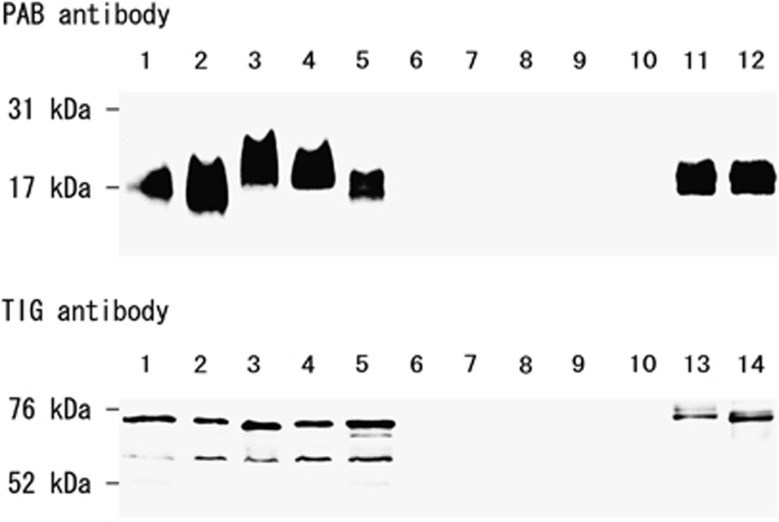
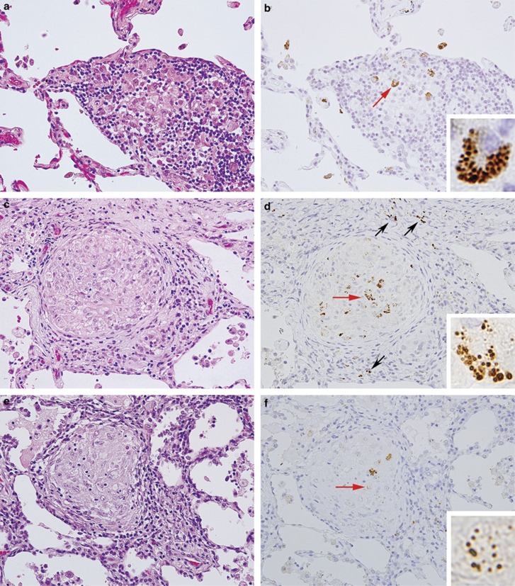
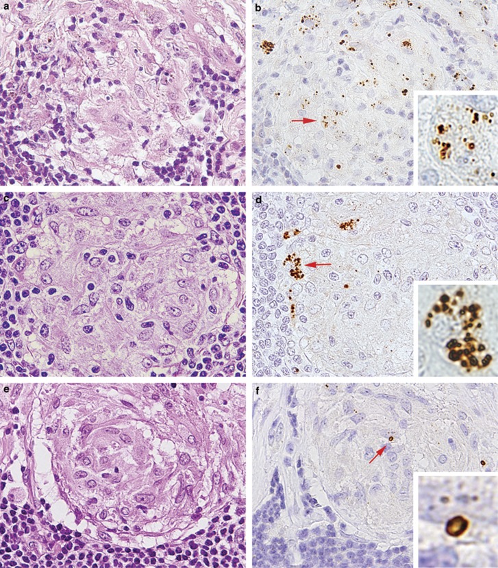
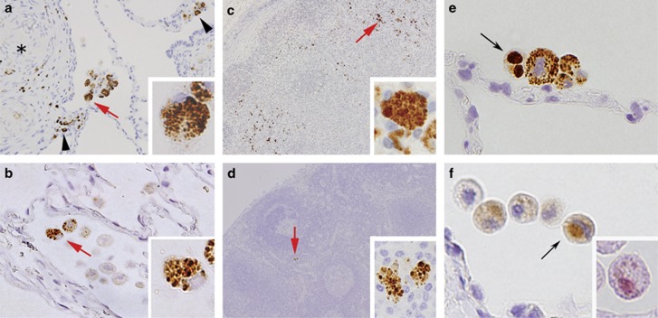
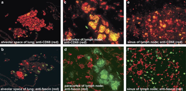
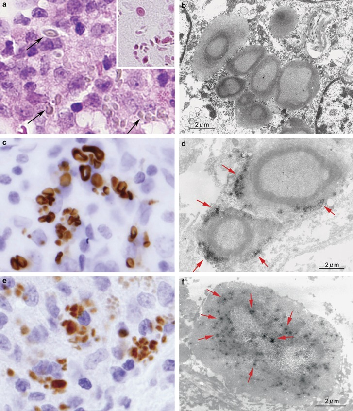
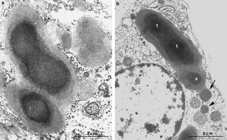
Sarcoidosis likely results from the exposure of a genetically susceptible subject to an environmental agent, possibly an infectious one. Mycobacterial and propionibacterial organisms are the most commonly implicated potential etiologic agents. Propionibacterium acnes is the only microorganism, however, found in sarcoid lesions by bacterial culture. To evaluate the pathogenic role of this indigenous bacterium, we screened for the bacterium in sarcoid and non-sarcoid tissues using immunohistochemical methods with novel P. acnes-specific monoclonal antibodies that react with cell-membrane-bound lipoteichoic acid (PAB antibody) and ribosome-bound trigger-factor protein (TIG antibody). We examined formalin-fixed and paraffin-embedded samples of lungs and lymph nodes from 196 patients with sarcoidosis, and corresponding control samples from 275 patients with non-sarcoidosis diseases. The samples were mostly from Japanese patients, with 64 lymph node samples from German patients. Immunohistochemistry with PAB antibody revealed small round bodies within sarcoid granulomas in 20/27 (74%) video-assisted thoracic surgery lung samples, 24/50 (48%) transbronchial lung biopsy samples, 71/81 (88%) Japanese lymph node samples, and 34/38 (89%) German lymph node samples. PAB antibody did not react with non-sarcoid granulomas in any of the 45 tuberculosis samples or the 34 samples with sarcoid reaction. In nongranulomatous areas, small round bodies detected by PAB antibody were found in alveolar macrophages of lungs and paracortical macrophages of lymph nodes from many sarcoid and some non-sarcoid patients. Large-spheroidal acid-fast bodies, Hamazaki-Wesenberg bodies, which were found in 50% of sarcoid and 15% of non-sarcoid lymph node samples, reacted with both PAB and TIG antibodies. Electron microscopy revealed that these Hamazaki-Wesenberg bodies had a single bacterial structure and lacked a cell wall with occasional protrusions from the body. The high frequency and specificity of P. acnes, detected by PAB antibody within sarcoid granulomas, indicates that this indigenous bacterium might be the cause of granuloma formation in many sarcoid patients.
Figures
Comment in
-
Histological differences between sarcoidosis and lung cancer-related sarcoid reaction.Respir Investig. 2020 Sep;58(5):421-424. doi: 10.1016/j.resinv.2020.04.003. Epub 2020 May 29. Respir Investig. 2020. PMID: 32482594 No abstract available.
References
-
- Hunninghake GW, Costabel U, Ando M, et al. ATS/ERS/WASOG statement on sarcoidosis. American Thoracic Society/European Respiratory Society/World Association of Sarcoidosis and other Granulomatous Disorders. Sarcoidosis Vasc Diffuse Lung Dis. 1999;16:149–173. - PubMed
-
- Baughman RP, Lower EE, du Bois RM. Sarcoidosis. Lancet. 2003;361:1111–1118. - PubMed
-
- Newman LS, Rose CS, Bresnitz EA, et al. A case control etiologic study of sarcoidosis: environmental and occupational risk factors. Am J Respir Crit Care Med. 2004;170:1324–1330. - PubMed
-
- McGrath DS, Goh N, Foley PJ, et al. Sarcoidosis: genes and microbes—soil or seed. Sarcoidosis Vasc Diffuse Lung Dis. 2001;18:149–164. - PubMed
-
- du Bois RM, Goh N, McGrath D, et al. Is there a role for microorganisms in the pathogenesis of sarcoidosis. J Intern Med. 2003;253:4–17. - PubMed
Publication types
MeSH terms
Substances
LinkOut - more resources
Full Text Sources
Molecular Biology Databases
