. 2012 Aug 7;12(15):2678-86.
doi: 10.1039/c2lc40235a. Epub 2012 May 17.

Integrated rapid-diagnostic-test reader platform on a cellphone

Affiliations

Integrated rapid-diagnostic-test reader platform on a cellphone

Onur Mudanyali et al. Lab Chip. .

Abstract

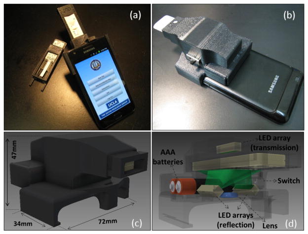
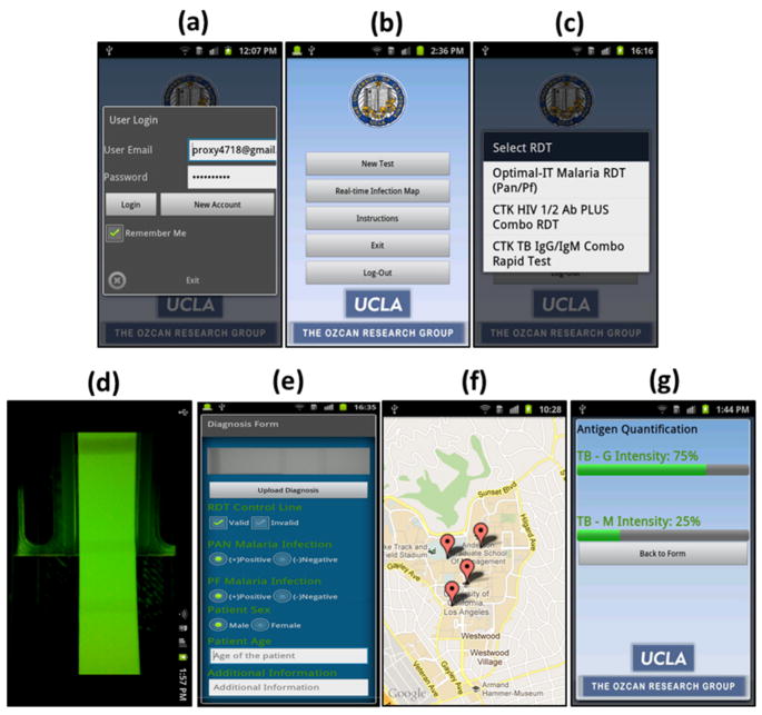
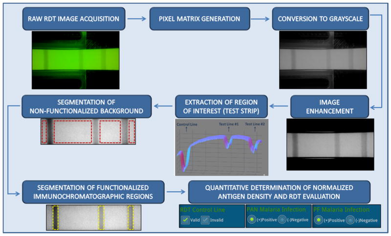
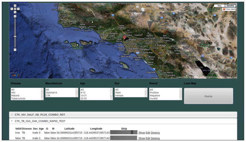
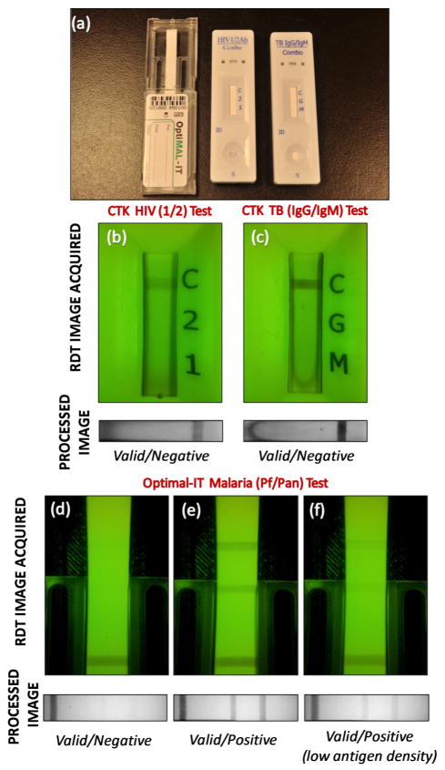
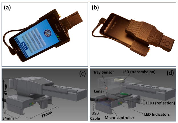
We demonstrate a cellphone-based rapid-diagnostic-test (RDT) reader platform that can work with various lateral flow immuno-chromatographic assays and similar tests to sense the presence of a target analyte in a sample. This compact and cost-effective digital RDT reader, weighing only ~65 g, mechanically attaches to the existing camera unit of a cellphone, where various types of RDTs can be inserted to be imaged in reflection or transmission modes under light-emitting diode (LED)-based illumination. Captured raw images of these tests are then digitally processed (within less than 0.2 s per image) through a smart application running on the cellphone for validation of the RDT, as well as for automated reading of its diagnostic result. The same smart application then transmits the resulting data, together with the RDT images and other related information (e.g., demographic data), to a central server, which presents the diagnostic results on a world map through geo-tagging. This dynamic spatio-temporal map of various RDT results can then be viewed and shared using internet browsers or through the same cellphone application. We tested this platform using malaria, tuberculosis (TB) and HIV RDTs by installing it on both Android-based smartphones and an iPhone. Providing real-time spatio-temporal statistics for the prevalence of various infectious diseases, this smart RDT reader platform running on cellphones might assist healthcare professionals and policymakers to track emerging epidemics worldwide and help epidemic preparedness.


Figures

Fig. 1
Fig. 1
(a–b) Different views of our smart RDT reader prototype installed on an Android phone (Samsung Galaxy S II). This light-weight (65 grams) and compact attachment can be repeatedly attached/detached to the cellphone body without the need for fine alignment and modification. To accommodate various RDT types using the same base attachment, customized sample trays are used. (c–d) Schematic diagrams of the designed optical RDT reader attachment are shown. It utilizes three LED arrays with diffusers to ensure uniform illumination of the tests which are loaded to the attachment with customized trays. Two of the LED arrays are located underneath the RDT tray to illuminate it for reflection imaging, whereas a third LED array is used for top illumination to record transmission images of the same RDT. Powered by two AAA batteries, reflection and transmission illumination LED arrays are controlled manually by a physical switch located on the side of the attachment or digitally through the audio jack of the cellphone device (if available). Depending on the format of the diagnostic test, users can switch between the two illumination schemes (reflection vs. transmission) to acquire an image of the RDT with high contrast. This image is then rapidly processed within less than 0.2 sec/image through a custom-developed application running on the cellphone (see Fig. 2) to generate an automated report that consists of test validation and reading of the diagnostic results as well as quantification of the test lines’ color intensities.
Fig. 2
Fig. 2
(a) User Login screen of the Android application running on the cell phone device that is shown in Fig. 1. (b) Main Menu of the application is shown. (c) Once user selects to image a new RDT, a pull-down menu of the already-configured RDTs pops up. (d) Upon the selection of RDT type and illumination scheme (reflection or transmission), application displays a raw image of the test. User can then touch the screen to capture and digitally process and evaluate the test results. (e) RDT Evaluation Menu displays test results including validity of the test (valid/invalid), decision of infection (positive/negative) for each test type as well as other information that the user can manually enter. The same application then uploads this test result to our database/server (f) that is protected by password. In case wireless connection is not available, it saves the results to the cellphone memory to be transmitted later. (g) Our application can also quantify the relative antigen density for each test line. To perform this antigen density measurement, RDT application needs to be calibrated by imaging a blank un-functionalized test (for minimum color intensity) and a saturated test (for maximum color intensity) that is functionalized using positive control antigens. Then the application returns the ratio of antigen density compared to reference values per test line. Note that for each RDT type, this calibration needs to be performed only once.
Fig. 3
Fig. 3
Block diagram of the automated image processing algorithm that is run by our cellphone based smart RDT reader application. In this case, a reflection image of the RDT is shown and processed.
Fig. 4
Fig. 4
Web interface of the Real Time RDT Map generated by our server is shown. This global database stores and organizes test results uploaded by users. The server displays the test data on an internet browser using Google Maps and can filter the data displayed based on several attributes, including: disease type, test location and time/date, RDT type/manufacturer, patient age, etc. It can be reached from the main menu of the cellphone application or from a remote location using a personal computer or other mobile devices running a web browser.
Fig. 5
Fig. 5
(a) Rapid-diagnostic-tests (RDTs) used in this study. Left: Optimal-IT P. falciparum-specific and Pan-specific Malaria Test. Middle: CTK HIV 1/2 Ab PLUS Combo Test. Right: CTK TB IgG/IgM Combo Test. (b) HIV 1/2 Combo RDT has a control reagent line indicating the validity of the test, and two pre-deposited antigen (HIV-1 and HIV-2) coated lines indicating the infections. (c) TB IgG/IgM Combo RDT is also a lateral-flow based immunoassay for simultaneous detection and differentiation of IgM anti-Mycobacterium Tuberculosis (M.TB) and IgG anti-M.TB in human serum or whole blood. Digitally processed reflection images of RDTs which are activated by fresh whole blood samples and their automated decisions are shown below the raw RDT images (b and c) acquired by our RDT reader platform. Optimal-IT Malaria RDT (d, e, and f) detects Plasmodium antigens (pLDH) using monoclonal antibodies. We imaged (in reflection) a malaria test that was activated using human blood (d) and was analyzed by our RDT application running on the cellphone. Moreover, we tested malaria RDTs using positive control wells which are previously coated by recombinant antigens of P. falciparum. In reflection mode, we imaged these RDTs that were activated using recommended malaria antigen densities (e), yielding a positive test result as expected, as well as using highly diluted antigens (f), i.e., beyond the recommended values. Refer to Figure 6 for further details.
Fig. 6
Fig. 6
Cross-sectional analysis of automatically processed malaria RDT images captured in reflection mode. The RDT reader shown in Fig. 1(a) was used to evaluate Malaria RDTs activated with 3 different batches of Positive Control Well Antigen (PCWA) at dilution levels of PCWA/20μl (1x), PCWA/40μl (2x), PCWA/60μl (3x). The above figure illustrates the mean of 10 pre-processed RDT image cross-sections (for each dilution level) and the standard deviation of maximum peak values of each test line. Mean and standard deviation values of the Control, Pan-Malaria, and P. Falciparum lines of the 1x dilution level are 8.37, 3.54, 6.02; 0.89, 0.77, and 0.72, respectively. Mean and standard deviation values of the Control, Pan-Malaria, and P. Falciparum lines of the 2x dilution level are 8.11, 1.29, 3.10; 2.10, 0.77, and 1.38, respectively. Mean and standard deviation values of the Control, Pan-Malaria, and P. Falciparum lines of the 3x dilution level are 7.62, 0.60, 2.03; 0.50, 0.22, and 0.51, respectively. Exemplary cellphone images of RDTs for each dilution level are also shown in the inset.
Fig. 7
Fig. 7
A smart RDT reader prototype (attached to Samsung Galaxy S II) which is entirely powered by the cell-phone battery through a USB connection. Controlled through a cost-effective micro-controller, two LED indicators are also used in this design to automatically detect (i) if the attachment is successfully powered through USB; and (ii) if the RDT is properly loaded into the reader and is ready to be imaged.

References

    1. Banoo S, Bell D, Bossuyt P, Herring A, Mabey D, et al. Nat Rev Microbio. 2006;4:21–31. - PubMed
    1. Vasoo S, Stevens J, Singh K. Clin Infect Dis. 2009;49:1090–1093. - PMC - PubMed
    1. Mills LA, Kagaayi J, Shott JP, Newell K, Bwanika JB, Ssempijja V, Aluma S, Quinn TC, Reynolds SJ, Gray RH. Trans R Soc Trop Med Hyg. 2010;104(3):237–239. - PMC - PubMed
    1. Wongsrichanalai C, Barcu MJ, Muth S, Sutamihardja A, Wernsdorfer WH. Am J Trop Med Hyg. 2007;77:119–127. - PubMed
    1. Dhorda M, Piola P, Nyehangane D, Tumwebaze B, Nalusaji A, Nabasumba C, Turyakira E, McGready R, Ashley E, Guerin PJ, Snounou G. Am J Trop Med Hyg. 2012;86(1):93–95. - PMC - PubMed

Publication types

MeSH terms