. 2012 Dec 15;520(18):4157-67.
doi: 10.1002/cne.23146.

Forebrain GABAergic projections from the dorsal raphe nucleus identified by using GAD67-GFP knock-in mice

Affiliations

Forebrain GABAergic projections from the dorsal raphe nucleus identified by using GAD67-GFP knock-in mice

Sun Jung Bang et al. J Comp Neurol. .

Abstract

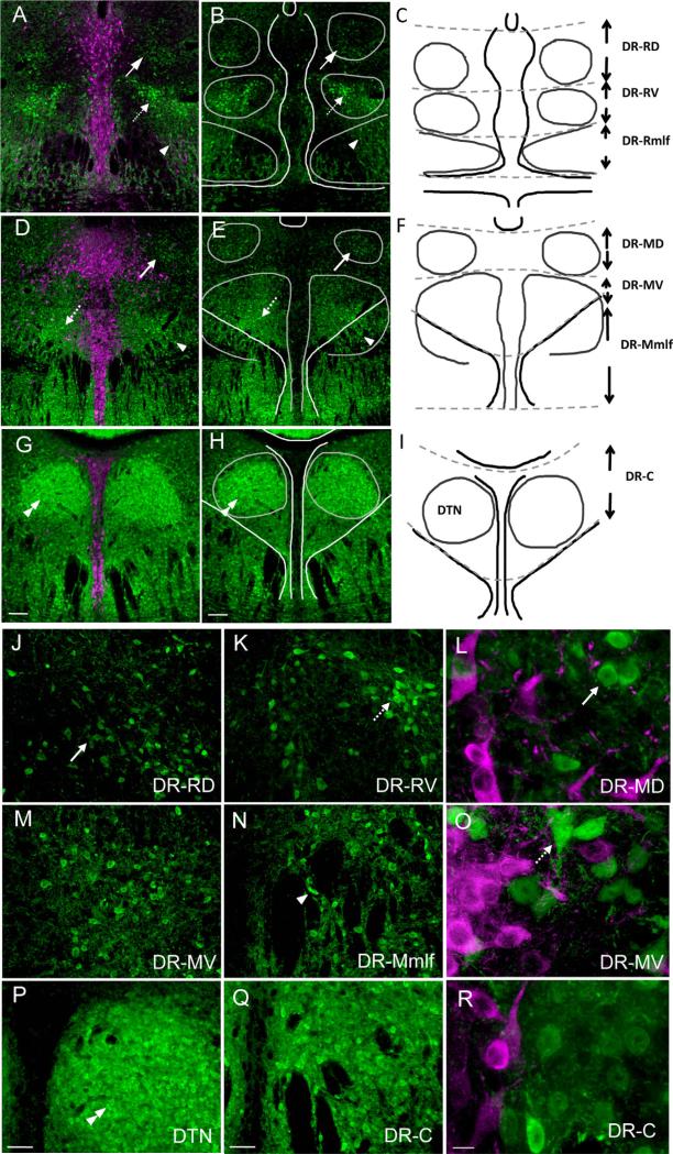
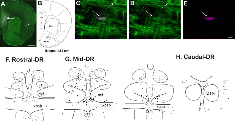
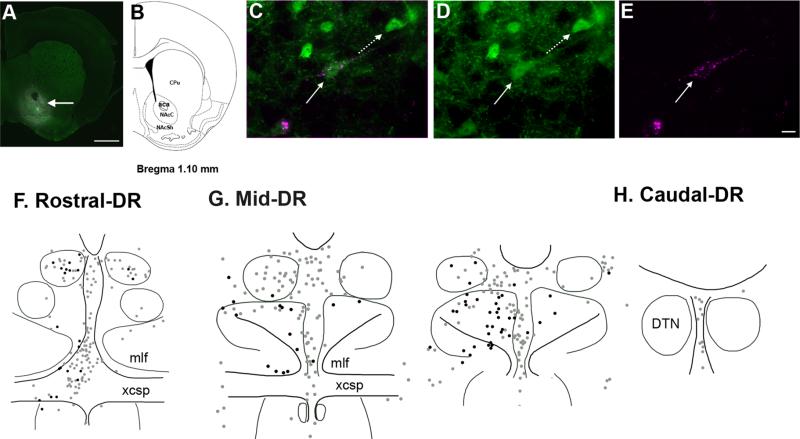
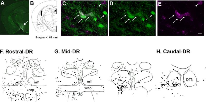
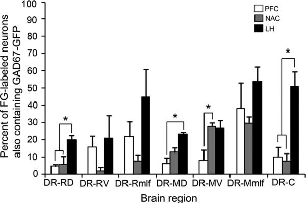
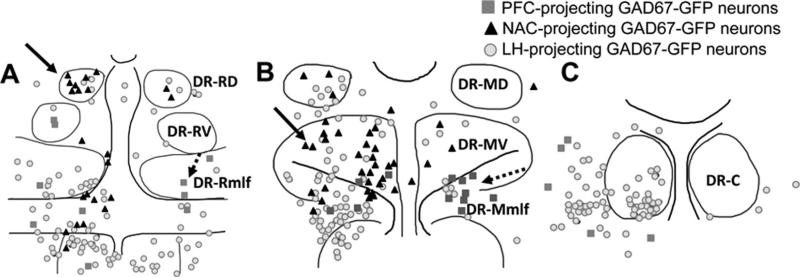
The dorsal raphe nucleus (DR) contains serotonergic (5-HT) neurons that project widely throughout the forebrain. These forebrain regions also receive innervation from non-5-HT neurons in the DR. One of the main groups of non-5-HT neurons in the DR is γ-aminobutyric acid (GABA)ergic, but their projections are poorly understood due to the difficulty of labeling these neurons immunohistochemically. To identify GABAergic projection neurons within the DR in the current study, we used a knock-in mouse line in which expression of green fluorescent protein (GFP) is controlled by the glutamic acid decarboxylase (GAD)67 promotor. Projections of GAD67-GFP neurons to the prefrontal cortex (PFC), nucleus accumbens (NAC), and lateral hypothalamus (LH) were evaluated by using retrograde tract tracing. The location of GAD67-GFP neurons projecting to each of these areas was mapped by rostrocaudal and dorsoventral location within the DR. Overall, 16% of DR neurons projecting to either the PFC or NAC were identified as GAD67-GFP neurons. GAD67-GFP neurons projecting to the PFC were most commonly found ventrally, in the rostral two-thirds of the DR. NAC-projecting GAD67-GFP neurons had an overlapping distribution that extended dorsally. GAD67-GFP neurons made a larger contribution to the projection of the DR to the LH, accounting for 36% of retrogradely labeled neurons, and were widespread throughout the DR. The current data indicate that DR GABAergic neurons not only may have the capacity to influence local network activity, but also make a notable contribution to DR output to multiple forebrain targets. J. Comp. Neurol. 520:4157-4167, 2012. © 2012 Wiley Periodicals, Inc.


Figures

Figure 1
Figure 1
Distribution of GFP-expressing GAD67 neurons (green) with respect to TPOH-stained neurons (magenta) in the DR. A,B: In the rostral third of the DR, a dorsal cluster of neurons dimly immunolabeled for GFP (arrows) and a ventral, more intensely labeled group (dashed arrows) are present. GAD67–GFP-immunolabeled neurons continue ventrally, interspersed within fiber bundles of the mlf (arrowhead). C: Reference schematic drawing showing landmarks used to plot the location of projecting GAD67–GFP neurons, with their abbreviations: DR-RD, rostral dorsal DR; DR-RV, rostral ventral DR; DR-Rmlf, rostral medial longitudinal fasciculus. D,E: In sections passing through the middle third of the DR, again dorsal (arrows) and ventral (dashed arrows) groups of GAD67–GFP neurons are seen, as well as those extending through the mlf (arrowhead). F: Reference schematic showing the middle dorsal (DR-MD), middle ventral (DR-MV), and middle medial longitudinal fasciculus (DR-Mmlf). G,H: In the caudal third of the DR, packed GAD67–GFP neurons are visible within the dorsal tegmental nucleus of Gudden (DTN, double arrowhead), as well as dispersed in the surrounding neuropil. I: Reference schematic drawing. J: GAD67-GFP neurons in the rostral and dorsal DR typically have small cell bodies (arrow). K: Rostral and ventral, GAD67–GFP neurons can be more intensely labeled and are larger (dashed arrow). L: Higher magnification of round, GAD67–GFP neurons (arrow) located dorsally. M: Many GAD67–GFP neurons located mid-ventral. N: GAD67–GFP neurons interspersed between fiber bundles (arrowhead). O: GAD67–GFP neurons (dashed arrow) with variable morphology in mid-ventral (DR-MV). P: GAD67–GFP neurons packed in the DTN (double arrowhead). Q: GAD67–GFP neurons dispersed caudally in the DR. R: Higher magnification of neurons located in the caudal DR. Scale bar = 100 μm in G (applies to A,D,G) and H (applies to B,E,H); 50 μm in P (applies to J,M,P) and Q (applies to K,N,Q); 10 μm in R (applies to L,O,R). [Color figure can be viewed in the online issue, which is available at wileyonlinelibrary.com.]
Figure 2
Figure 2
Location of neurons projecting to the prefrontal cortex (PFC), with and without GAD67–GFP in the DR. A,B: A representative image of FG injection in the PFC (arrow) with corresponding bran atlas section adapted from Paxinos and Franklin (2001). Drug diffusion was restricted within the prelimbic (PrL) and infralimbic cortex (IL). C–E: Immunofluorescence labeling for GFP (green) and FG (magenta) shows a double-labeled GAD67–GFP neurons (straight arrow) retrogradely labeled from the PFC. In addition, a neuron solely labeled for GFP is visible (dashed arrow) in the mlf region of the DR. D and E show immunolabeling for GFP and FG, respectively; they are merged in C. F–H: Schematic illustrations of brain sections showing distribution of PFC-projecting neurons that are either GAD67–GFP (black dots) or non-GAD67–GFP (gray dots) at the rostral, middle, and caudal levels of the DR. Abbreviations: Cg1, cingulate cortex; CPu, caudate putamen; DTN, the dorsal tegmental nuclei of Gudden; mlf, medial longitudinal fasciculus; NAcC, nucleus accumbens core; NAcSh, nucleus accumbens shell; xcsp, decussation of the superior cerebral peduncle. For other abbreviations, see list. Scale bar = 1 mm in A; 20 micro-meter in E (applies to C–E). [Color figure can be viewed in the online issue, which is available at wileyonlinelibrary.com.]
Figure 3
Figure 3
Distribution of DR neurons projecting to the nucleus accumbens (NAC), with and without GAD67–GFP. A,B: A representative image showing FG injection in the NAC with corresponding brain atlas section adapted from Paxinos and Franklin (2001). FG diffused into both the core (NAcC) and shell (NAcSh) regions of the NAC (arrow) surrounding the anterior commissure (aca). C–E: An example of NAC-projecting GAD67–GFP neurons (straight arrow) double labeled for GFP (green) and FG (magenta). An adjacent GAD67–GFP neuron (dashed arrow) lacks immunolabeling for FG. Immunolabeling for GFP and FG observed individually in D and E, respectively. F–H: Schematic illustrations of the distribution of NAC-projecting neurons that contain GAD67–GFP (black dots) or lack GAD67–GFP (gray dots) in the rostral, middle, and caudal DR. For abbreviations, see list. Scale bar = 1 mm in A; 20 micrometer in E (applies to C–E). [Color figure can be viewed in the online issue, which is available at wileyonlinelibrary.com.]
Figure 4
Figure 4
Distribution of neurons projecting to the lateral hypothalamus (LH) with and without GAD67–GFP. A,B: FG diffusion was restricted in the LH (arrow) as shown in the representative image with corresponding brain atlas section adapted from Paxinos and Franklin (2001). C–E: GAD67–GFP neurons projecting to the LH (straight arrows) were double labeled with both GFP (green) and FG (magenta), whereas other GAD67–GFP neurons not projecting to the LH (dashed arrow) are labeled with GFP only. A neuron projecting to the LH lacking GAD67–GFP (arrowhead) is also visible. F–H: Schematic illustration showing the locations of LH-projecting GAD67–GFP (black dots) and non-GAD67–GFP neurons (gray dots) in the rostral, middle, and caudal levels of the DR, respectively. Abbreviations: Am, amygdala; HP, hippo-campus; LH, lateral hypothalamus. For other abbreviations, see list. Scale bar = 1 mm in A; 20 micrometer in E (applies to C–E). [Color figure can be viewed in the online issue, which is available at wileyonlinelibrary.com.]
Figure 5
Figure 5
Mean percentage of the total numbers of neurons projecting to each target region that were double immunolabeled for GAD67–GFP, calculated for each subdivision of the DR examined. Mean percentage of GAD67–GFP neurons in each subdivision projecting to the PFC, NAC, or LH are denoted in white, gray, and black bars, respectively. Asterisks denote significant group differences. For abbreviations, see list.
Figure 6
Figure 6
Schematic illustrations showing the locations of GAD67–GFP neurons projecting to each target region examined at the rostral (A), mid (B), and caudal (C) levels of the DR. Within both the rostral (A) and middle third of the DR (B), NAC-projecting GAD67–GFP neurons (black triangles, arrows) tended to be located dorsal to PFC-projecting GAD67–GFP neurons (dark gray squares, dashed arrows) that were mostly located in the mlf region. LH-projecting GAD67–GFP neurons (light gray circles) were distributed throughout the DR subdivisions. At the caudal level (C), GAD67–GFP neurons primarily projected to the LH. For abbreviations, see list.

References

    1. Ansorge MS, Hen R, Gingrich JA. Neurodevelopmental origins of depressive disorders. Curr Opin Pharmacol. 2007;7:8–17. - PubMed
    1. Bassett JP, Tullman ML, Taube JS. Lesions of the tegmentomammillary circuit in the head direction system disrupt the head direction signal in the anterior thalamus. J Neurosci. 2007;27:7564–7577. - PMC - PubMed
    1. Brown RE, McKenna JT, Winston S, Basheer R, Yanagawa Y, Thakkar MM, McCarley RW. Characterization of GABAergic neurons in rapid-eye-movement sleep controlling regions of the brainstem reticular formation in GAD67-green fluorescent protein knock-in mice. Eur J Neurosci. 2008;27:352–363. - PMC - PubMed
    1. Bubar MJ, Stutz SJ, Cunningham KA. 5-HT(2C) receptors localize to dopamine and GAD67–GFP neurons in the rat mesoaccumbens pathway. PLoS One. 2011;6:e20508. - PMC - PubMed
    1. Chang B, Daniele CA, Gallagher K, Madonia M, Mitchum RD, Barrett L, Vezina P, McGehee DS. Nicotinic excitation of serotonergic projections from dorsal raphe to the nucleus accumbens. J Neurophysiol. 2011;106:801–808. - PMC - PubMed

Publication types

MeSH terms

Substances