. 2012 May 18;336(6083):925-31.
doi: 10.1126/science.1215317.

Multiple spectral inputs improve motion discrimination in the Drosophila visual system

Affiliations

Multiple spectral inputs improve motion discrimination in the Drosophila visual system

Trevor J Wardill et al. Science. .

Abstract

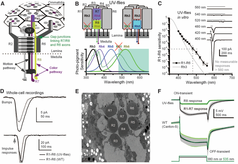
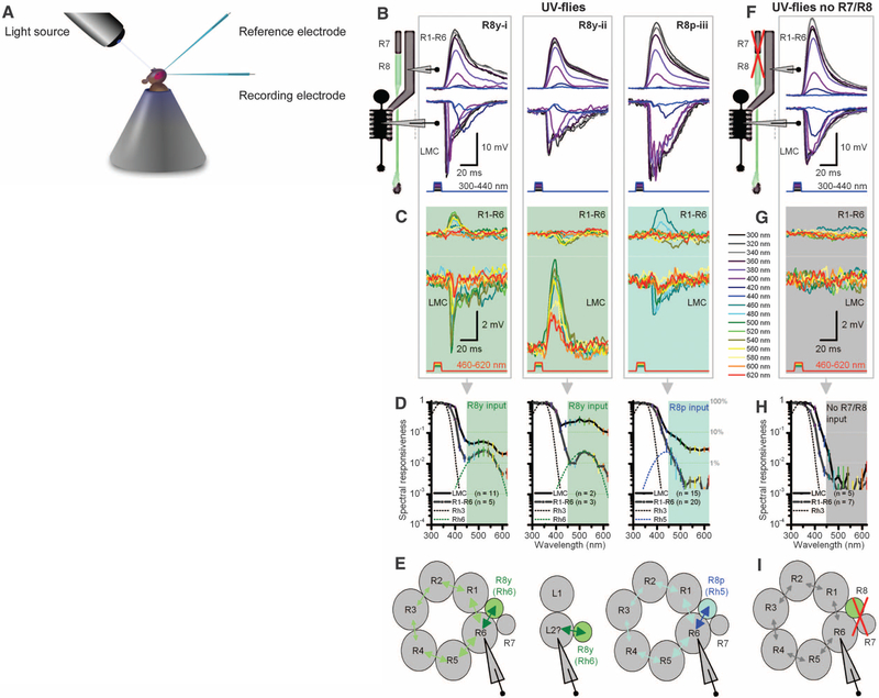
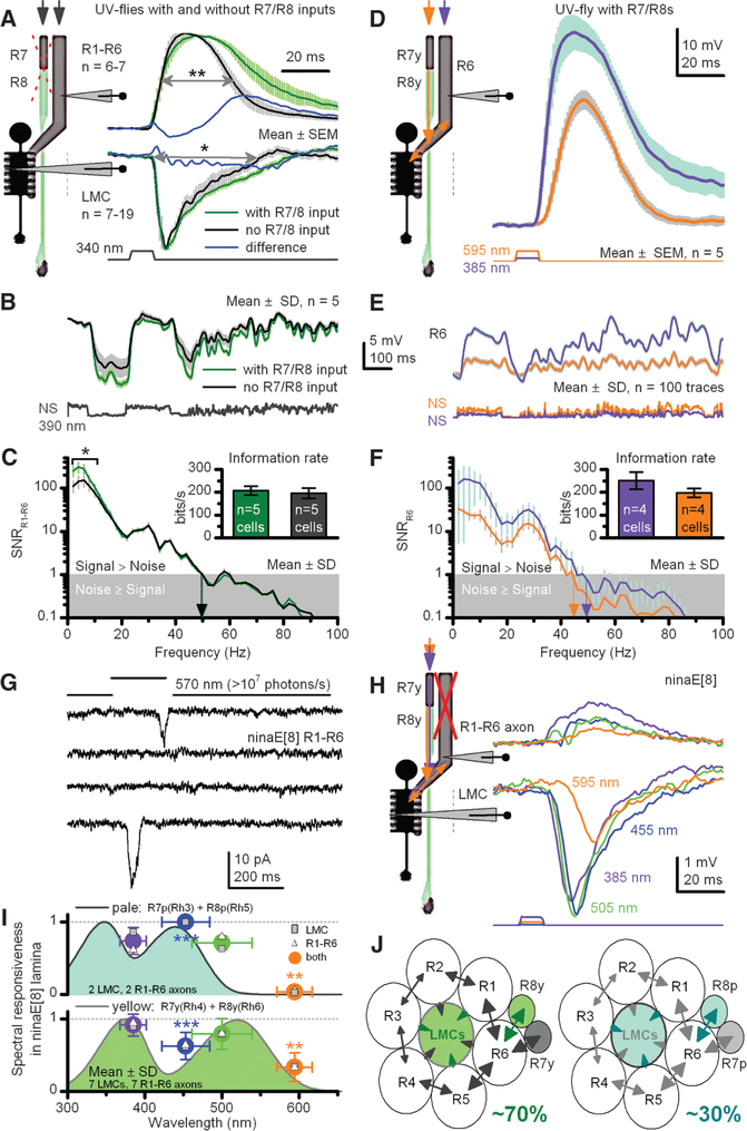
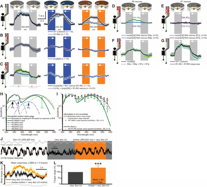
Color and motion information are thought to be channeled through separate neural pathways, but it remains unclear whether and how these pathways interact to improve motion perception. In insects, such as Drosophila, it has long been believed that motion information is fed exclusively by one spectral class of photoreceptor, so-called R1 to R6 cells; whereas R7 and R8 photoreceptors, which exist in multiple spectral classes, subserve color vision. Here, we report that R7 and R8 also contribute to the motion pathway. By using electrophysiological, optical, and behavioral assays, we found that R7/R8 information converge with and shape the motion pathway output, explaining flies' broadly tuned optomotor behavior by its composite responses. Our results demonstrate that inputs from photoreceptors of different spectral sensitivities improve motion discrimination, increasing robustness of perception.


Figures

Fig. 1.
Fig. 1.
Manipulating spectral sensitivity of the motion pathway to elucidate circuit computations. (A)Schematic offly photo-receptors innervating motion and color pathways. A light point is sampled by six outer photoreceptors (R1–R6) in neighboring ommatidia and by central R7/R8 photoreceptors. These R1–R6s inner-vate LMCs (L1 and L2) in the first optic neuropile, lamina, whereas R7/R8 synapseinthesecondneuropile, medulla. Gap junctions link R7/R8 to R6 just before R1–R6 are brought together (superpositioned) for synaptic transmission (28). (B) In UV flies, UV-sensitive Rh3-opsin is expressed in R1–R6s, which contain nonfunctional Rh1-opsin (ninaE8). This circumvents the spectral overlap between motion andR8pathways.(C)Spectral sensitivity of light-induced currents (inset) in dissociated R1–R6s of UV flies, diminishing <1/100,000 at ≥480 nm (mean ± SD; four photoreceptors). (Inset) Example responses to 2-ms flashes (arrows) or 50- or 500-ms stimuli (bars). (D) Although single-photon responses (bumps) can be larger than those of WT (Canton-S) and impulse responses last slightly longer, their responses are very similar to their WT counterparts, indicating normal-like phototransduction (fig. S4, A and B) (35). (E) Photoreceptors of UV flies have normal-like ultrastructure. (F) Electroretinograms (ERGs) show comparable dynamics to those of WT flies: Preferred colors evoke large receptor components and on and off transients, indicating normal synaptic transmission from R1–R6 to LMCs (means T SD; six flies). With UV flies, one can separate the responses for green (R8s) and UV (R1–R7s).
Fig. 2.
Fig. 2.
Motion pathway receives information from R8 photo-receptors in color pathway. (A) Schematic of in vivo recordings using monochromatic light. (B) Three intracellular response types from R1–R6 photoreceptors and postsynaptic LMCs to subsaturating short-wavelength (300 to 420 nm) pulses of equal energy. (C) Stimulating the same representative cells (B, i to iii) with equal long-wavelength (420 to 620 nm) pulses, which are undetectable by Rh3, reveals unexpected responses both in polarity and spectral responsiveness. (D) Spectral responsiveness of R1–R6 and LMC outputs confirms a second peak or bulge, which matches the sensitivity of R8y (Rh6) or R8p (Rh5) photoreceptors; right graph also includes cells with subtle long-wavelength range extensions. (E) Results suggest a model (29) in which information spreads via gap junctions from each R8 to R6 axon, and further to other photoreceptors in the same cartridge, before transmission to LMCs. Depolarizing responses (420 to 620 nm) in some LMCs suggest gap junctions between L2 and R8 axons. (F) In UV flies in which R1–R8 phototransduction was deactivated (norpA36) and then rescued only in R1–R6 (i.e., light-insensitive R7/R8), R1–R6 and LMC have a sensitivity to short-wavelength stimuli that is similar to when R7/R8 are functional (B). (G) These R1–R6s and LMCs cannot detect 460 to 620 nm. (H) Their spectral responsiveness matches the Rh3 sensitivity of dissociated cells (Fig. 1C). (I) With light-insensitive R7/R8s, information processing occurs independently within the motion pathway.
Fig. 3.
Fig. 3.
R7/R8 photoreceptors transmit information to a motion pathway shaping R1–R6 and LMC outputs. (A) Normalized R1–R6 and LMC outputs to a saturating UV impulse in UV flies with or without light-sensitive R7/R8s. Wider responses suggest extra R7 inputs. Differences highlight the change over time between the genotypes. (B) R1–R6 outputs to naturalistic UV-intensity series; NS, with or without light-sensitive R7/R8s. (C) Corresponding signal-to-noise ratios (SNRs) and information transfers (inset). (D) Apart from the three response types in Fig. 2C, ~10% of R1–R6s from UV flies also responded strongly (>25 mV) to amber, signifying direct R8y input to R6 (29) (figs. S6 and S7). (E) Responses to UV and amber NS [different from (B)] in one presumptive R6. (F) High SNR with amber responses, containing ~70% of information of maximal UV responses (inset, three R6s). (G) Whole-cell recordings reveal that ninaE8 R1–R6s are profoundly light-insensitive (fig. S3B), lacking macroscopic light-induced current; ultrabright green-yellow light rarely evokes single-photon responses (two shown). (H) An in vivo example where presynaptic ninaE8 R1–R6 axon and postsynaptic LMC generate small responses, following R7/R8s’ sensitivity, here R7y/R8y; similar recordings from nine LMCs and nine R1–R6s (fig. S5C). (I) Responsiveness of ninaE8 LMCs and R1–R6 axons track R7/R8-nomogram pairs. (J) Input to each lamina cartridge comes from R7, R8, R7y/R8y, or R7p/R8p pairs through gap junctions to R6 axons to drive synaptic output to LMCs. *P < 0.05; **P < 0.01, ***P < 0.005, one-way ANOVA.
Fig. 4.
Fig. 4.
Signals from R1–R6 and R7/R8 sum to generate optomotor responses. Responses (yaw torque) of tethered flying Drosophila to different color-field rotations (black-white, black-blue, and black-amber) to left and right. (A) Both UV flies (R1–R6s expressing Rh3-opsin) and WT respond to broad spectral range of motions. a.u., arbitrary unit. (B) Mutant ninaE8-flies, having light-insensitive R1–R6 photoreceptors (Fig. 3G; motion pathway) but normal R7/R8 photo-receptors, react to an equally broad spectral range of motions as in (A), but with weaker responses. (C) norpA36 flies, having R1–R6 photoreceptors rescued with Rh1 or Rh3 opsins, react to broad spectral range of motions but with weaker and faster responses than in (A). (D) Rescuing light sensitivity only in R8y or R8p photo-receptors enables visual motion perception. (E) Rescuing light sensitivity only in R7y or R7p photoreceptors enables weaker motion perception (fig. S11B). (F) Responses of norpA36 flies with rescued light sensitivity in R7 and R8 photoreceptors equal the mean responses of ninaE8 flies. (G) Responses of norpA36 flies with rescued R1–R6 photoreceptors and ninaE8 flies (with normal R7/R8s) sum up the mean responses of WT flies. In (A) to (G), mean T SEM is shown in same scale. (H) Motion-vision range estimated by weighting R1–R8 inputs with their relative optomotor response strengths: (D) to (G). (I) Range coincides with LMCs’ spectral sensitivity. (J) UV motion is dimmed until a fly’s optomotor responses nearly cease. Adding amber light strengthens responses. (K) Corresponding averages. (L) R8y input improves motion discrimination by ~36% (P = 0.0004; t test for mean > 1).

References

    1. Nishida S, Watanabe J, Kuriki I, Tokimoto T, Curr. Biol 17, 366 (2007). - PubMed
    1. Takeuchi T, De Valois KK, Hardy JL, Vision Res. 43, 1159 (2003). - PubMed
    1. Barlow HB, Behav. Brain Sci 24, 602 (2001). - PubMed
    1. Knill DC, Pouget A, Trends Neurosci. 27, 712 (2004). - PubMed
    1. Schiller PH, Proc. Natl. Acad. Sci. U.S.A 107, 17087 (2010). - PMC - PubMed

Publication types

MeSH terms