

Neonatal lupus syndrome in a Nigerian child
- PMID: 22605870
- PMCID: PMC3369365
- DOI: 10.1136/bcr.01.2012.5710
Neonatal lupus syndrome in a Nigerian child
Abstract
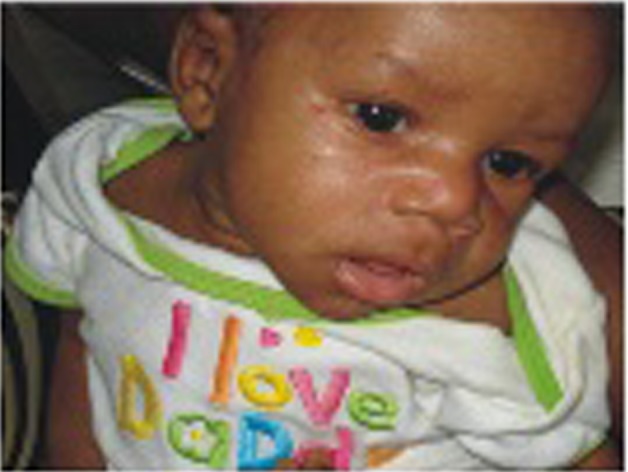
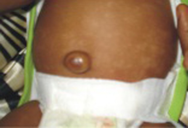
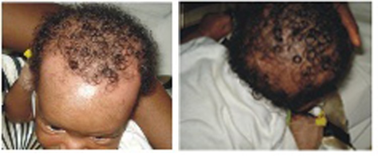
Neonatal lupus is a rare syndrome resulting from passively transferred maternal autoantibodies during pregnancy. A male infant was delivered at term to a 29-year-old primiparous woman who was diagnosed of systemic lupus erythematosus 2 years earlier and had detectable levels of autoantibodies (antinuclear antibody (ANA), anti-dsDNA, anti-Ro and anti-La/SSB) in second trimester. However, the pregnancy was otherwise uneventful. He presented at the age of 8 week with a widespread hypopigmented macular rash on the trunk and patchy alopecia involving the hair line and the occipito-parietal regions of 3 weeks duration, anaemia and symptomatic thrombocytopaenia. Serologic test for autoantibodies was positive for ANA and anti-La/SSB. Further evaluation was normal. He was managed conservatively with blood products and topical corticosteroids. Mother was also advised to avoid direct exposure to sunlight and fluorescent light. Haematological parameters gradually normalised over 2 months and the skin lesions resolved completely by the age of 6 months.
Conflict of interest statement
Figures
Similar articles
-
Seeing double: annular diaper rash in twins.Pediatr Dermatol. 2015 May-Jun;32(3):410-3. doi: 10.1111/pde.12508. Epub 2015 Jan 30. Pediatr Dermatol. 2015. PMID: 25639144
-
Neonatal lupus with atypical cardiac and cutaneous manifestation.BMJ Case Rep. 2013 Jul 8;2013:bcr2013009249. doi: 10.1136/bcr-2013-009249. BMJ Case Rep. 2013. PMID: 23839605 Free PMC article.
-
Rowell Syndrome in Nigeria: Systemic Lupus Erythematosus Presenting as Recurrent Erythema Multiforme in a Young Woman.Acta Dermatovenerol Croat. 2019 Sep;27(3):200-201. Acta Dermatovenerol Croat. 2019. PMID: 31542069
-
[Neonatal lupus syndrome: Literature review].Rev Med Interne. 2015 Mar;36(3):159-66. doi: 10.1016/j.revmed.2014.07.013. Epub 2014 Sep 17. Rev Med Interne. 2015. PMID: 25240481 Review. French.
-
Anti-RO/SSA and anti-La/SSB antibodies: Association with mild lupus manifestations in 645 childhood-onset systemic lupus erythematosus.Autoimmun Rev. 2017 Feb;16(2):132-135. doi: 10.1016/j.autrev.2016.12.004. Epub 2016 Dec 14. Autoimmun Rev. 2017. PMID: 27988434 Review.
Cited by
-
The Child as a Surrogate for Diagnosis of Lupus in the Mother.Case Rep Rheumatol. 2017;2017:8247591. doi: 10.1155/2017/8247591. Epub 2017 Mar 13. Case Rep Rheumatol. 2017. PMID: 28386505 Free PMC article.
-
Neonatal lupus presenting as a non-specific rash in primary care.BMJ Case Rep. 2020 Dec 13;13(12):e237463. doi: 10.1136/bcr-2020-237463. BMJ Case Rep. 2020. PMID: 33318248 Free PMC article.
References
-
- Tseng CE, Buyon JP. Neonatal lupus syndromes. Rheum Dis Clin North Am 1997;23:31–54 - PubMed
-
- Lee LA, Frank MB, McCubbin VR, et al. Autoantibodies of neonatal lupus erythematosus. J Invest Dermatol 1994;102:963–6 - PubMed
-
- Adelowo OO, Oguntona SA. Pattern of systemic lupus erythematosus among Nigerians. Clin Rheumatol 2009;28:699–703 - PubMed
-
- Ho SY, Esscher E, Anderson RH, et al. Anatomy of congenital complete heart block and relation to maternal anti-Ro antibodies. Am J Cardiol 1986;58:291–4 - PubMed
-
- Clancy RM, Backer CB, Yin X, et al. Genetic association of cutaneous neonatal lupus with HLA class II and tumor necrosis factor alpha: implications for pathogenesis. Arthritis Rheum 2004;50:2598–603 - PubMed
Publication types
MeSH terms
Substances
Supplementary concepts
LinkOut - more resources
Full Text Sources
Medical
Research Materials