. 2012;13(4):4920-4936.
doi: 10.3390/ijms13044920. Epub 2012 Apr 19.

Structural and functional characterization of two alternative splicing variants of mouse Endothelial Cell-Specific Chemotaxis Regulator (ECSCR)

Affiliations

Structural and functional characterization of two alternative splicing variants of mouse Endothelial Cell-Specific Chemotaxis Regulator (ECSCR)

Wen Wu et al. Int J Mol Sci. 2012.

Abstract

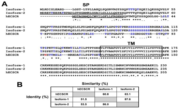
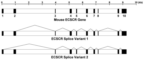
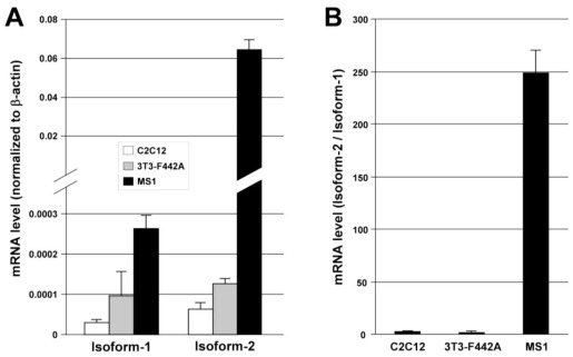
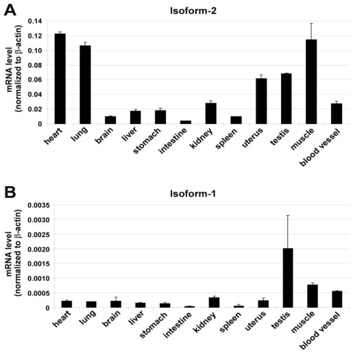
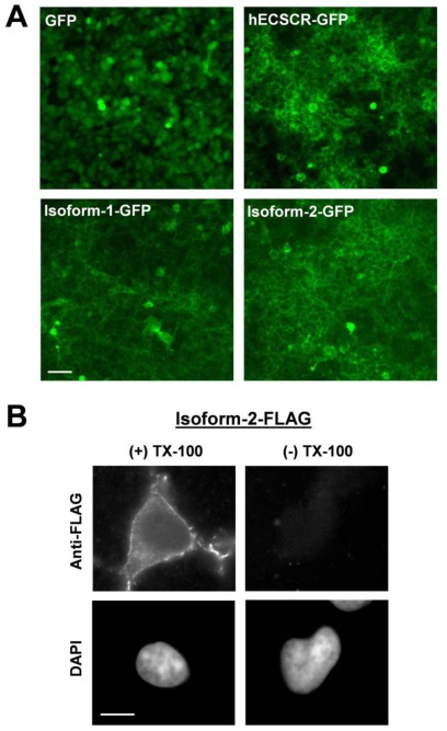
Endothelial cells (ECs) that line the lumen of blood vessels are important players in blood vessel formation, and EC migration is a key component of the angiogenic process. Thus, identification of genes that are specifically or preferentially expressed in vascular ECs and in-depth understanding of their biological functions may lead to discovery of new therapeutic targets. We have previously reported molecular characterization of human endothelial cell-specific molecule 2 (ECSM2)/endothelial cell-specific chemotaxis regulator (ECSCR). In the present study, we cloned two mouse full-length cDNAs by RT-PCR, which encode two putative ECSCR isoform precursors with considerable homology to the human ECSCR. Nucleotide sequence and exon-intron junction analyses suggested that they are alternative splicing variants (ECSCR isoform-1 and -2), differing from each other in the first and second exons. Quantitative RT-PCR results revealed that isoform-2 is the predominant form, which was most abundant in heart, lung, and muscles, and moderately abundant in uterus and testis. In contrast, the expression of isoform-1 seemed to be more enriched in testis. To further explore their potential cellular functions, we expressed GFP- and FLAG-tagged ECSCR isoforms, respectively, in an ECSCR deficient cell line (HEK293). Interestingly, the actual sizes of either ECSCR-GFP or -FLAG fusion proteins detected by immunoblotting are much larger than their predicted sizes, suggesting that both isoforms are glycoproteins. Fluorescence microscopy revealed that both ECSCR isoforms are localized at the cell surface, which is consistent with the structural prediction. Finally, we performed cell migration assays using mouse endothelial MS1 cells overexpressing GFP alone, isoform-1-GFP, and isoform-2-GFP, respectively. Our results showed that both isoforms significantly inhibited vascular epidermal growth factor (VEGF)-induced cell migration. Taken together, we have provided several lines of experimental evidence that two mouse ECSCR splicing variants/isoform precursors exist. They are differentially expressed in a variety of tissue types and likely involved in modulation of vascular EC migration. We have also defined the gene structure of mouse ECSCR using bioinformatics tools, which provides new information towards a better understanding of alternative splicing of ECSCR.

Keywords: ECSCR/ECSM2; alternative splicing; cDNA cloning and expression; endothelial cell migration; exon-intron boundary; gene structure; isoform.


Figures

Figure 1
Figure 1
RT-PCR to obtain full-length cDNAs encoding mouse ECSCR isoform-1 and isoform-2 precursors. Total RNAs were extracted from mouse large blood vessels (aorta) and RT-PCR was performed to obtain full-length cDNAs that encode mouse ECSCR isoform-1 (A) and isoform-2 (B), respectively. DNA ladders are shown.
Figure 2
Figure 2
Identification of mouse ECSCR isoform-1 and isoform-2 precursors. (A) Amino acid sequence alignment of mouse ECSCR isoform-1, isoform-2, and human ECSCR precursors. Putative signal peptides (SP) are underlined and a single transmembrane domain (TM) is boxed. Predicted N-glycosylation and O-glycosylation sites are shown in red and blue, respectively. (B) Percentages of identity and similarity among the amino acid sequences of mouse ECSCR isoform-1, isoform-2, and human ECSCR precursors.
Figure 3
Figure 3
Gene structure of mouse ECSCR and alternative splicing events. The reconstructed mouse ECSCR gene is based on new data presented in this study. A total of ten exons are indicated by closed boxes.
Figure 4
Figure 4
Expression of ECSCR isoform-1 and isoform-2 in cultured mouse cell lines. (A) The mRNA levels of isoform-1 and isoform-2 measured by qRT-PCR. C2C12, mouse myoblasts; 3T3-F442A, mouse preadipocytes; MS1, mouse endothelial cells. (B) The ratio of mRNA level of isoform-2 to that of isoform-1 indicates that isoform-2 is the predominant form expressed in endothelial MS1 cells. Data are mean ± SD (n = 3).
Figure 5
Figure 5
Expression of ECSCR isoform-1 and isoform-2 in a variety of mouse tissues. The mRNA levels of isoform-2 (A) and isoform-1 (B) measured by qRT-PCR. Blood vessels examined here were large vessels (aorta). Data are mean ± SD (n = 3).
Figure 6
Figure 6
Heterologous expression of mouse ECSCR isoform-1 and isoform-2 proteins suggests that they are likely glycoproteins. (A and B) GFP- or FLAG-tagged human (h) ECSCR, mouse isoform-1, and isoform-2 were expressed I HEK293 cells, respectively. The expression of fusion proteins was confirmed by Western blot (WB) analysis with anti-GFP (A) or anti-FLAG (B) antibody. The glycosylated (mature) forms of fusion proteins are indicated by asterisks. (C) Comparison of the actual molecular weights (MW) of the fusion proteins in kilodaltons (kDa) on SDS-PAGE with their predicted molecular weights (MW) based amino acid sequences.
Figure 7
Figure 7
Cell surface localization of mouse ECSCR isoforms. (A) HEK293 cells expressing GFP alone, human (h) ECSCR-GFP, mouse ECSCR isoform-1-GFP, and isoform-2-GFP, respectively, were visualized by fluorescent microscopy. Scale bar, 50 μm. (B) Mouse ECSCR isoform-2 tagged with FLAG at its C-terminus was expressed in HEK293 cells and the transfectants were costained with anti-FLAG antibody and DAPI. The cell surface localization of ECSCR-FLAG was detected only in the cells that were permeabilized with 0.1% Triton X-100. Scale bar, 20 μm.
Figure 8
Figure 8
Overexpression of mouse ECSCR isoforms in MS1 cells inhibits endothelial cell motility. MS1 cells overexpressing GFP alone, mouse ECSCR isoform-1-GFP, and isoform-2-GFP, respectively, were subjected to Transwell migration assay, in which VEGF (10 ng/mL) was used as a chemoattractant as detailed in Methods. The cells were allowed to migrate for 5 h. The number of migrated cells per imaging field for each condition was counted. Data are mean ± SD (n = 10), * p < 0.05, ** p < 0.01.

References

    1. Risau W. Mechanisms of angiogenesis. Nature. 1997;386:671–674. - PubMed
    1. Carmeliet P., Jain R.K. Angiogenesis in cancer and other diseases. Nature. 2000;407:249–257. - PubMed
    1. Folkman J., D’Amore P.A. Blood vessel formation: What is its molecular basis? Cell. 1996;87:1153–1155. - PubMed
    1. Jacobs J. Combating cardiovascular disease with angiogenic therapy. Drug Discov. Today. 2007;12:1040–1045. - PubMed
    1. Rahimi N. Vascular endothelial growth factor receptors: Molecular mechanisms of activation and therapeutic potentials. Exp. Eye Res. 2006;83:1005–1016. - PMC - PubMed

Publication types

Substances

LinkOut - more resources