Comparative Study
. 2012;7(5):e36659.
doi: 10.1371/journal.pone.0036659. Epub 2012 May 11.

Differential actions of chlorhexidine on the cell wall of Bacillus subtilis and Escherichia coli

Affiliations
Comparative Study

Differential actions of chlorhexidine on the cell wall of Bacillus subtilis and Escherichia coli

Hon-Yeung Cheung et al. PLoS One. 2012.

Abstract

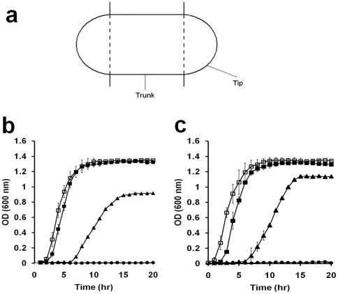
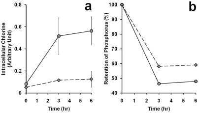
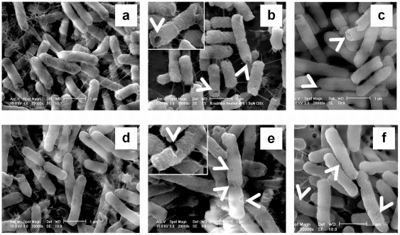
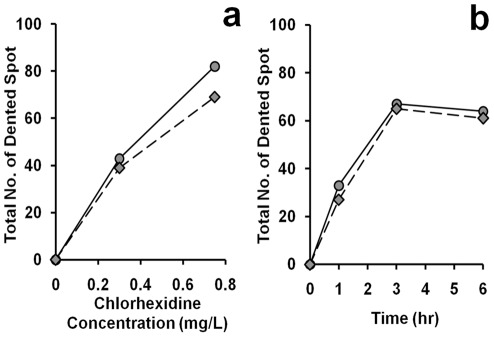
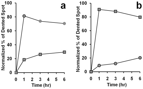
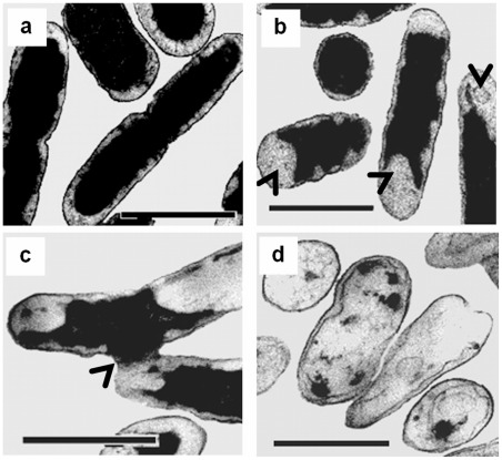
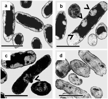
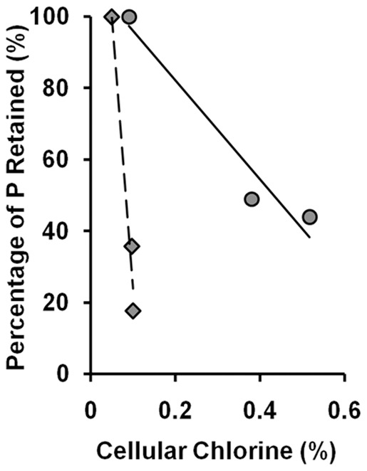
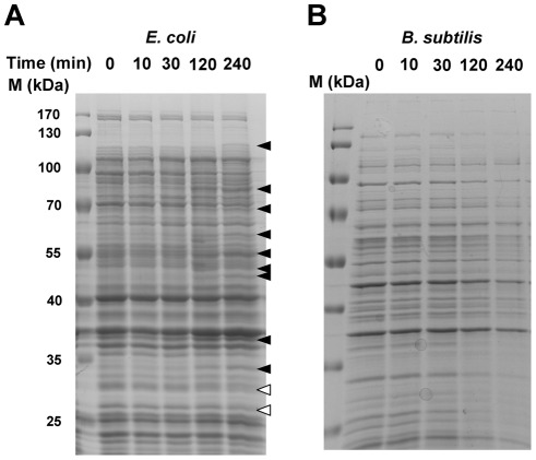
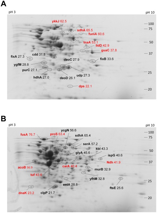
Chlorhexidine is a chlorinated phenolic disinfectant used commonly in mouthwash for its action against bacteria. However, a comparative study of the action of chlorhexidine on the cell morphology of gram-positive and gram-negative bacteria is lacking. In this study, the actions of chlorhexidine on the cell morphology were identified with the aids of electron microscopy. After exposure to chlorhexidine, numerous spots of indentation on the cell wall were found in both Bacillus subtilis and Escherichia coli. The number of indentation spots increased with time of incubation and increasing chlorhexidine concentration. Interestingly, the dented spots found in B. subtilis appeared mainly at the hemispherical caps of the cells, while in E. coli the dented spots were found all over the cells. After being exposed to chlorhexidine for a prolonged period, leakage of cellular contents and subsequent ghost cells were observed, especially from B subtilis. By using 2-D gel/MS-MS analysis, five proteins related to purine nucleoside interconversion and metabolism were preferentially induced in the cell wall of E. coli, while three proteins related to stress response and four others in amino acid biosynthesis were up-regulated in the cell wall materials of B. subtilis. The localized morphological damages together with the biochemical and protein analysis of the chlorhexidine-treated cells suggest that chlorhexidine may act on the differentially distributed lipids in the cell membranes/wall of B. subtilis and E. coli.


Conflict of interest statement

Competing Interests: The authors have declared that no competing interests exist.

Figures

Figure 1
Figure 1. Effect of chlorhexidine on the growth of bacterial cells.
(a) Definition of trunk and tip of a bacterial cell. The two hemispherical caps are the tips while the middle cylindrical zone is the trunk of a rod-shaped bacterial cell as described in Materials and methods. (b and c) The growth of B. subtilis (b) and E. coli (c) was monitored by measuring the optical density of the culture. The culture was sampled at a particular time point and OD at 600 nm was measured. The concentrations of chlorhexidine added to the culture were: 0 (open rectangle), 0.25 (filled rectangle), 0.5 (closed triangle), 0.75 (closed circle) mg/L. Each plot of the curve is the average of three readings and standard deviation is shown.
Figure 2
Figure 2. Uptake of chlorine and loss of phosphorus by bacterial cells in 0.75
mg/L chlorhexidine. The cellular contents of chlorine (a) and phosphorus (b) in the culture of B. subtilis (diamond) or E. coli (circle) at different time points were subjected to analysis using EDAX with an environmental scanning electron microscope (ESEM) as described in Materials and Methods. Standard errors were calculated based on the results from three separate samples. Experiment in (b) was repeated twice and the averages of the two were plotted.
Figure 3
Figure 3. Morphological changes of B. subtilis and E. coli after exposure to chlorhexidine and acetone.
The two species of bacterial cells exposed to 0.75 mg/L of chlorhexidine for 4 h were harvested and prepared as described in Materials and Methods. They were examined with an environmental scanning electron microscope after glutaraldehyde fixation. From (a) to (c) the cells were B. subtilis and from (d) to (f) the cells were E. coli. B. subtilis (a) and E. coli (d) cells were directly mounted for electron-microscopic examination without any CHX treatment. In (b) and (e), the cells were treated with 0.75 mg/L chlorhexidine for 4 h. For (c) and (f), the cells were treated with 5% and 7% acetone for 4 h, respectively. These results were confirmed in two independent experiments. Magnification in all electron micrographs was x 20,000. Bar  = 1 μm. Magnification of the inserts of (b) and (e) was x 30,000.
Figure 4
Figure 4. Total number of dented spot observed on cell surface.
The dented spots on both the trunk and tips were counted in 50 cells of B. subtilis (diamond) and E. coli (circle) from cultures with (a) different chlorhexidine concentrations for 6 hr and (b) treated with 0.75 mg/L of chlorhexidine for different lengths of time. These results were confirmed in two independent experiments.
Figure 5
Figure 5. Time course of chlorhexidine treatment on the distribution of dented spots on the cell surface.
Bacterial cells were incubated in media with 0.75 mg/L chlorhexidine over a 6 hr period. The bacterial cells at a particular time point were harvested and examined under an environmental scanning electron microscope (ESEM). The number of dented spots at the trunk (rectangle) and tip (circle) was counted in B. subtilis (a) and E. coli (b). Each time point represents the average number of dented spot on 50 cells. These results were confirmed in two independent experiments.
Figure 6
Figure 6. Cytological changes of B. subtilis after treatment with chlorhexidine.
Control cells possess intact cell membrane, cell wall, and complete cell content (a). After 4 h of incubation with 0.75 mg/L chlorhexidine, treated cells showed detached cytoplasmic membrane from the cell wall at the two poles of a rod cell (arrows in b), leakage of cell content (arrow in c) and formation of ghost cells (d). These results were confirmed in two independent experiments. Magnification in all cases  = ×20,500. Bar  = 1 μm.
Figure 7
Figure 7. Cytological changes of E. coli after treated with chlorhexidine.
Control cells possess intact cell membrane, cell wall, and complete cell content (a). After 4 h of incubation with 0.75 mg/L chlorhexidine, the treated E. coli cells showed detached cytoplasmic membrane from the cell wall at both poles and cylindrical part of the cells (see arrows in b), leakage of cell content (see arrow in c) and formation of ghost cells (d). These results were confirmed in two independent experiments. Magnification in all cases  = ×20,500. Bar  = 1 μm.
Figure 8
Figure 8. Effect of chlorhexidine exposure on phospholipids content in B. subtilis and E. coli.
B. subtilis (diamond) and E. coli (circle) were treated with 0, 0.3, and 0.75 mg/L of chlorhexidine for 3 h and then subjected to EDAX to determine the contents of phosphorus and chlorine retained in the cells. The results were confirmed in two independent experiments.
Figure 9
Figure 9. Time course of CHX treatment on the total protein contents in both species of bacteria.
SDS polyacrylamide gel separation of the total cellular proteins from (A) E. coli and (B) B. subtilis samples treated with CHX at different time points. The black arrow heads indicate proteins that were induced by the CHX treatment and hollow arrow head indicates protein that was suppressed in expression after the CHX treatment. The experiments were repeated twice and one is shown here.
Figure 10
Figure 10. 2D gel of cell wall/membrane proteins from E. coli and B. subtilis after CHX treatment.
Proteins extracted from the cell wall fraction of (A) E. coli and (B) B. subtilis were separated by isoelectric focusing (pH 3–10) and subsequent 12% SDS polyacrylamide gel followed by Coomassie Blue staining. Protein spots that increased in intensity were circled with black solid lines and those with decreased in intensity were circled with broken lines. The numbers on the left were the the molecular weight of protein markers in kDa. The pI of the protein along the first dimension was shown on the top of the gel with pH 3 on the left and pH 10 on the right. The identity of the protein spots was labelled with their gene name and their molecular weight. The experiments were repeated twice and showed similar spot patterns.

References

    1. McBain AJ, Bartolo RG, Catrenich CE, Charbonneau D, Ledder RG, et al. Effects of a chlorhexidine gluconate-containing mouthwash on the vitality and antimicrobial susceptibility of in vitro oral bacterial ecosystems. Appl Environ Microbiol. 2003;69:4770–4776. - PMC - PubMed
    1. Sreenivasan PK, Gittins E. The effects of a chlorhexidine mouthrinse on culturable microorganisms of the tongue and saliva. Microbiol Res. 2004;159:365–370. - PubMed
    1. Kanisavaran ZM. Chlorhexidine gluconate in endodontics: an update review. Int Dent J. 2008;58:247–257. - PubMed
    1. Ellepola ANB, Samaranayake LP. Adjunctive use of chlorhexidine in oval candidoses: a review. Oral Dis. 2001;7:11–17. - PubMed
    1. al-Tannir MA, Goodman HS. A review of chlorhexidine and its use in special populations. Spec Care Dentist. 1994;14:116–122. - PubMed

Publication types

MeSH terms

LinkOut - more resources