. 2012;7(5):e37035.
doi: 10.1371/journal.pone.0037035. Epub 2012 May 14.

Peptide array X-linking (PAX): a new peptide-protein identification approach

Affiliations

Peptide array X-linking (PAX): a new peptide-protein identification approach

Hirokazu Okada et al. PLoS One. 2012.

Abstract

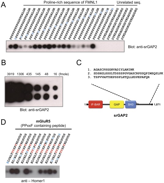
Many protein interaction domains bind short peptides based on canonical sequence consensus motifs. Here we report the development of a peptide array-based proteomics tool to identify proteins directly interacting with ligand peptides from cell lysates. Array-formatted bait peptides containing an amino acid-derived cross-linker are photo-induced to crosslink with interacting proteins from lysates of interest. Indirect associations are removed by high stringency washes under denaturing conditions. Covalently trapped proteins are subsequently identified by LC-MS/MS and screened by cluster analysis and domain scanning. We apply this methodology to peptides with different proline-containing consensus sequences and show successful identifications from brain lysates of known and novel proteins containing polyproline motif-binding domains such as EH, EVH1, SH3, WW domains. These results suggest the capacity of arrayed peptide ligands to capture and subsequently identify proteins by mass spectrometry is relatively broad and robust. Additionally, the approach is rapid and applicable to cell or tissue fractions from any source, making the approach a flexible tool for initial protein-protein interaction discovery.


Conflict of interest statement

Competing Interests: The authors have declared that no competing interests exist.

Figures

Figure 1
Figure 1. Experimental outline of PAX methodology.
(A) Arrays of bait peptides are synthesized onto PEG-based membrane supports. The photo-activatable amino acid cross-linker (pBpa) is incorporated into the bait peptides so that each peptide spot has pBpa at a different position in the sequence (see inset schematic). (B, C) The membrane is incubated with cell lysate and subjected to 350–365 nm light to cross-link with interacting proteins. (D) The indirect and non-specific interactors are removed by high stringent, denaturing washes. (E, F) Each strip of the bait peptide spots is cut off the membrane, further chopped into small pieces, and trypsinized. (G, H) The samples are filtered to remove PEG and subjected to LC-MS/MS analysis to identify the photo-trapped proteins.
Figure 2
Figure 2. PAX-captured proteins are MS identifiable.
(A) PAX captures a known interacting protein following over-expression in FreeStyle 293 cells. An array of peptides consisting of a proline-rich sequence of FMNL1 and a control sequence were incubated with FreeStyle 293 cell lysate overexpressing srGAP2, photo-crosslinked and washed at high stringency. Immunoblotting with srGAP2 antibody detected phototrapping of srGAP2 by PAX. pBpa in the bait peptides is indicated in blue. (B) Comparative immunoblotting of srGAP2 antigen dot-blot. Different amounts of srGAP2 were spotted onto nitrocellulose membrane and simultaneously immunoblotted with srGAP2 antibody. (C) Identification of srGAP2 by mass spectrometry. The five spots with the highest amount of crosslinked srGAP2 from a duplicate blot of (A) were subjected to MS/MS. 3 unique peptides (underlined) covering 8% of the entire sequence of srGAP2 protein were identified. (D) PAX captures a known interacting protein from mouse brain lysate. Peptide array comprising mGluR5 sequences with a PPXXF motif was incubated with mouse brain lysate, photo-crosslinked and subjected to high stringency washes. Immunoblotting with Homer1 antibody indicates photo-trapping of Homer1 by PAX. pBpa in the bait peptides is indicated in blue.
Figure 3
Figure 3. PAX successfully identifies interacting proteins from tissue lysates.
(A) Cluster analysis of MS/MS-identified proteins. Candidate interactors with the bait peptides from SOS1, Stonin2, Epsin1 and mGluR5 were hierarchically clustered using unbiased Pearson correlation of the mean normalized spectral counts. Protein clusters of single bait interactors are indicated with blue bars. (B) Identification of selective bait interactors. Single bait interactors identified in (A) are exhibited. Proteins in brown are known to bind to the corresponding bait peptides. Bioinformatics analysis revealed that proteins in orange were previously shown to interact with the bait proteins. (C) Protein interaction domain filtering. Proteins that showed association to the control bait peptide were eliminated from the MS-identified proteins, and the rest was subjected to cluster analysis. Subsequently, the proteins that contain domains anticipated to interact with consensus binding motifs found in bait peptides were selected as high confidence interactors. (D) Identification of the interactors with anticipated peptide recognition domains. Proteins were colored as explained in (B).
Figure 4
Figure 4. PAX identifies novel interacting proteins with WAVE1.
(A) Identification of ligands for a WAVE1 derived peptide. MS/MS data for WAVE1 trapped proteins from brain lysate were combined with those for control, SOS1, Stonin2, Epsin1 and mGluR5 and subjected to cluster analysis as in Fig. 3A. Vasp and Gas7 are known to interact with other members of the WAVE/WASP family proteins. (B) Protein interaction domain filtering. Enah and Amph, in addition to Vasp and Gas7, were identified as WAVE1 peptide-interacting proteins. (C) Only 3 of the 13 proteins identified in (B) were identified as abundant proteins using label-free LC/MS/MS quantification of mouse brain lysates. (D) WAVE1 peptide interactome. PAX identified high confidence interactions (red dotted arrows) with WAVE1. The previously known interactions are indicated with black arrows. The WAVE1 selective binding proteins contain domains (shown in red) that are expected to interact with the WAVE1 polyproline peptide (see Table 1). (E–F) Co-immunoprecipitation assays with full-length WAVE1 and (E) Amph or (F) Gas7. (G) GST pulldown of Enah using the proline peptide bait of WAVE1. (H) Co-immunoprecipitation of GFP-Enah with full-length WAVE1 and WAVE1 lacking the bait proline peptide. (I) Physiological interaction between WAVE1 and Enah. Reciprocal co-immunoprecipitation experiments were performed using rat brain extract (RBE). The precipitates were analyzed by Western blotting. (J) Co-localization of WAVE1 and Enah in fibroblast cells. Immunostaining of endogenous WAVE1 showed co-localization with Enah in MVD7 cells expressing GFP-tagged Enah.

References

    1. Pawson T, Nash P. Assembly of cell regulatory systems through protein interaction domains. Science. 2003;300:445–452. - PubMed
    1. Yu JW, Lemmon MA. Genome-wide analysis of signaling domain function. Current opinion in chemical biology. 2003;7:103–109. - PubMed
    1. Ponting CP, Russell RR. The natural history of protein domains. Annual review of biophysics and biomolecular structure. 2002;31:45–71. - PubMed
    1. Castagnoli L, Costantini A, Dall'Armi C, Gonfloni S, Montecchi-Palazzi L, et al. Selectivity and promiscuity in the interaction network mediated by protein recognition modules. FEBS letters. 2004;567:74–79. - PubMed
    1. Takenawa T, Suetsugu S. The WASP-WAVE protein network: connecting the membrane to the cytoskeleton. Nat Rev Mol Cell Biol. 2007;8:37–48. - PubMed

Publication types

MeSH terms

Substances