. 2012;7(5):e37169.
doi: 10.1371/journal.pone.0037169. Epub 2012 May 14.

MicroRNA regulation of human protease genes essential for influenza virus replication

Affiliations

MicroRNA regulation of human protease genes essential for influenza virus replication

Victoria A Meliopoulos et al. PLoS One. 2012.

Abstract

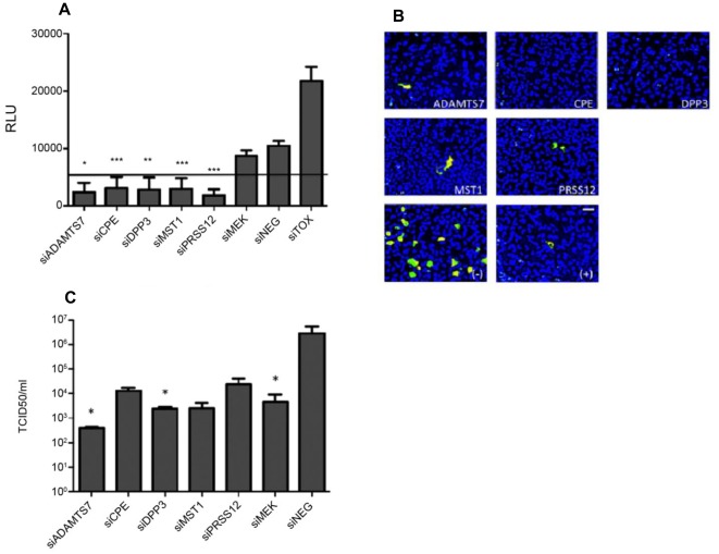
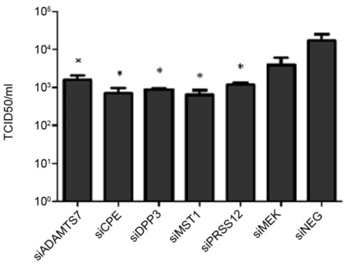
Influenza A virus causes seasonal epidemics and periodic pandemics threatening the health of millions of people each year. Vaccination is an effective strategy for reducing morbidity and mortality, and in the absence of drug resistance, the efficacy of chemoprophylaxis is comparable to that of vaccines. However, the rapid emergence of drug resistance has emphasized the need for new drug targets. Knowledge of the host cell components required for influenza replication has been an area targeted for disease intervention. In this study, the human protease genes required for influenza virus replication were determined and validated using RNA interference approaches. The genes validated as critical for influenza virus replication were ADAMTS7, CPE, DPP3, MST1, and PRSS12, and pathway analysis showed these genes were in global host cell pathways governing inflammation (NF-κB), cAMP/calcium signaling (CRE/CREB), and apoptosis. Analyses of host microRNAs predicted to govern expression of these genes showed that eight miRNAs regulated gene expression during virus replication. These findings identify unique host genes and microRNAs important for influenza replication providing potential new targets for disease intervention strategies.


Conflict of interest statement

Competing Interests: The authors have declared that no competing interests exist.

Figures

Figure 1
Figure 1. RNAi of 5 host protease genes down-regulated influenza virus replication.
A: A549 cells were reverse transfected with 50 nM of siRNA (SMARTpool) specific for the indicated genes (ADAMTS7, CPE, DPP3, MST1, PRSS12). After 48 hours, cytotoxicity was determined by adenylate kinase (AK) release. Cells treated with the siTOX control were considered 100% cytotoxic and all values were normalized to siTOX. RLU = relative luciferase units. * p<0.05 vs siNEG, ** p<0.01 vs siNEG, ***p<0.005 vs siNEG; siTOX vs all samples: p<0.001 (not shown). Line shows 20% of siTOX control. B: A549 cells were reverse transfected with 50 nM of siRNA (SMARTpool) specific for the indicated genes (ADAMTS7, CPE, DPP3, MST1, PRSS12). After 48 hours, cells were infected with A/WSN/33 at an MOI of 0.001. 48 hours post-infection, cells were fixed in 4% formaldehyde and stained with an anti-NP (green) monoclonal antibody followed by counterstain with DAPI (blue.) Positive (+) control: siMEK, negative (−) control: siNEG. Magnification is 20× (bar is 100 microns). C: Cells were transfected with 100 nM of a novel siRNA targeting a different seed site from the SMARTpool used in the primary screen and infected as in B. After 48 hours of infection, cellular supernatant was tested for infectious virus production by a modified TCID50. Data is expressed as TCID50/ml. Data is representative of two independent experiments. (*p<0.05 vs siNEG).
Figure 2
Figure 2. RNAi of individual host protease genes down-regulates replication of a clinical influenza isolate.
A549 cells were reverse transfected with 100 nM of the novel siRNA targeting siADAMTS7, siCPE, siDPP3, siMST1, and siPRSS12. After 48 hours, cells were infected with A/New Caledonia/20/99 at an MOI of 0.1 in the presence of 1 ug/ml TPCK-trypsin. After 48 hours of infection, cellular supernatant was tested for infectious virus production by a modified TCID50. Data is expressed as TCID50/ml. Data is representative of two independent experiments. (*p<0.05 vs. siNEG).
Figure 3
Figure 3. Analysis of host gene involvement in major cellular pathways.
A: A549 cells were reverse cotransfected with CRE/CREB reporter plasmid or the appropriate control plasmid and 100 nM of the novel siRNA. After 24 h incubation, the transfection media was replaced with culture media. Cells were mock infected or infected with A/WSN/33 at an MOI of 0.001 the following day. After 24 h, culture supernatant was analyzed for luciferase expression. Luciferase units were normalized to Renilla expression. * p<0.05, ** p<0.01 compared to siNEG control (mock), # p<0.05, ## p<0.01 compared to siNEG control (A/WSN/33) B: A549 cells were reversed cotransfected with NF-κB reporter plasmid or the appropriate control plasmid and 100 nM of the novel siRNA. After 24 h incubation, the transfection media was replaced with culture media. Cells were mock infected or infected with A/WSN/33 at an MOI of 0.001 the following day. After 24 h, culture supernatant was analyzed for luciferase expression. Luciferase units were normalized to Renilla expression. * p<0.05, ** p<0.01 compared to siNEG control (mock), # p<0.05, ## p<0.01 compared to siNEG control (A/WSN/33). Data is representative of three independent experiments.
Figure 4
Figure 4. RNAi of DPP3 (siDPP3) inhibits influenza replication by modulation of apoptotic genes.
A549 cells were reverse transfected with 50 nM of siDPP3 or siNEG. After 48 hours, cells were infected with A/WSN/33 at an MOI of 0.001. After 18 hours of infection, cellular RNA was isolated and apoptosis gene expression profiles were determined by array. Gene expression was normalized to GAPDH levels. Silencing DPP3 resulted in upregulated levels of the pro-apoptotic genes BCL2L10, TNFSF10, TNFSF25 and TNFSF8. Data is representative of three independent experiments. * p<0.05.
Figure 5
Figure 5. Effect of miRNA inhibition on host protease gene expression 24 h post-miRNA inhibitor treatment.
Host cell miRNAs of interest were evaluated for their effect on host gene hits by qPCR. A549 cells were treated with the appropriate miRNA inhibitor (25 nM) for 24 hours. Cellular RNA was isolated 24 hpi and evaluated by qPCR for host gene expression using a SYBRgreen assay with gene-specific primers. Gene expression was compared to cells transfected with siNEG (for siRNA) or NEG (non-targeting miRNA inhibitor) at the equivalent concentration. Data is normalized to GAPDH expression. miRNAs indicated on the x-axis refer to inhibition of those miRNAs. A: ADAMTS7 expression levels, B: CPE, C: DPP3, D: MST1, E: PRSS12. Data is representative of two independent experiments. (*p<0.05 versus siRNA treatment.).
Figure 6
Figure 6. Effect of miRNA inhibition on influenza replication.
A549 cells were treated with the appropriate miRNA inhibitor (25 nM) for 48 hours, followed by infection with A/WSN/33 (MOI = 0.001). Cellular supernatant was tested for infectious virus production by a modified TCID50 48 hpi. Data is expressed as TCID50/ml and is representative of two independent experiments.

References

    1. Kandel R, Hartshorn KL. Prophylaxis and treatment of influenza virus infection. BioDrugs. 2001;15:303–323. - PubMed
    1. Thompson WW, Shay DK, Weintraub E, Brammer L, Cox N, et al. Mortality associated with influenza and respiratory syncytial virus in the United States. JAMA. 2003;289:179–186. - PubMed
    1. Hale BG, Randall RE, Ortin J, Jackson D. The multifunctional NS1 protein of influenza A viruses. J Gen Virol. 2008;89:2359–2376. - PubMed
    1. Basler CF. Influenza viruses: basic biology and potential drug targets. Infect Disord Drug Targets. 2007;7:282–293. - PubMed
    1. Betakova T. M2 protein-a proton channel of influenza A virus. Curr Pharm Des. 2007;13:3231–3235. - PubMed

Publication types

MeSH terms