

Case Reports
doi: 10.1155/2011/679751.
Epub 2012 Jan 31.
Macular hole progression after intravitreal bevacizumab for hemicentral retinal vein occlusion
Affiliations
- PMID: 22606470
- PMCID: PMC3350047
- DOI: 10.1155/2011/679751
Item in Clipboard
Case Reports
Macular hole progression after intravitreal bevacizumab for hemicentral retinal vein occlusion
Case Rep Ophthalmol Med.
2011.
Abstract
Macular edema secondary to retinal vein occlusion is commonly being treated with off-label intravitreal bevacizumab with good outcomes. A significant reduction in macular edema and improvement in visual acuity is seen following such a treatment with no serious adverse effects. In the reported case, a full-thickness macular hole was noticed one month after intravitreal bevacizumab for macular edema secondary to hemicentral retinal vein occlusion. On a detailed review of the pre- and postoptical coherence tomography scans, it was realized that there was a preexisting stage 2-3 macular hole which was masked by the hemorrhages and edema at the fovea and the macular hole had progressed following the injection.
Figures
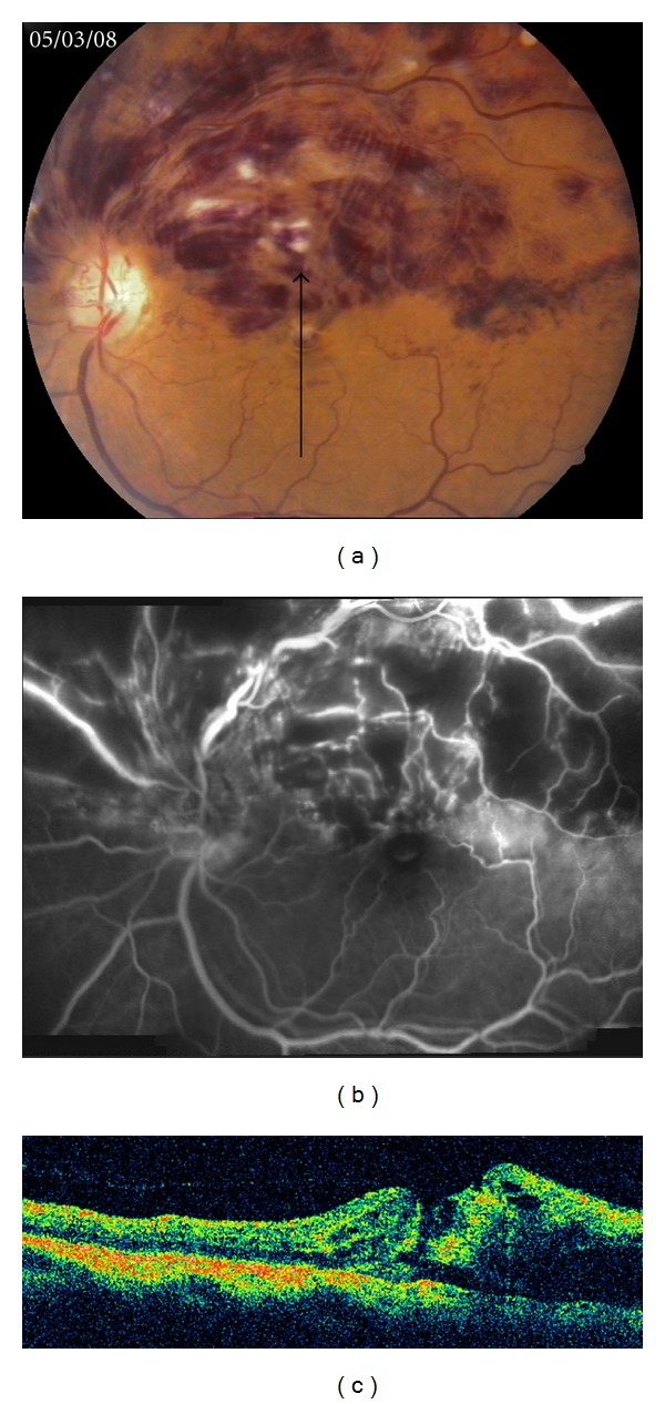
(a) Fundus photograph of the left eye showing superficial retinal hemorrhages, cotton wool spots, and tortuous and dilated retinal veins in the superior half of the retina involving the macula. These findings were consistent with a superior HCRVO. (b) Fluorescein angiography demonstrated blocked fluorescence due to retinal hemorrhages with areas of capillary non perfusion. (c) Vertical 5 mm OCT scan along the arrow shows cystoid edema with retinal hemorrhages in the superior half of the fovea (right half of the scan) and some retinal discontinuity which in retrospect was a stage 2-3 macular hole. Hyaloidal attachment is also seen.
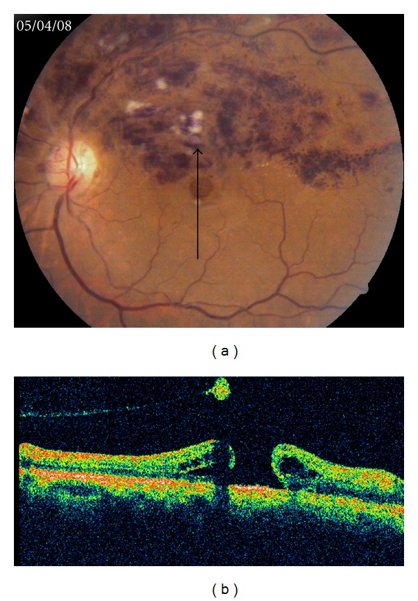
(a) Fundus photograph, one month after intravitreal bevacizumab injection, showing decreased haemorrhages with a full-thickness macular hole. (b) Vertical 5 mm OCT scan along the same line shows progression to a full-thickness macular hole with a detached hyaloid and a pseudooperculum causing a shadowing behind it.
References
-
- Pai SA, Shetty R, Vijayan PB, et al. Clinical, anatomic, and electrophysiologic evaluation following intravitreal bevacizumab for macular edema in retinal vein occlusion. American Journal of Ophthalmology. 2007;143(4):601–606. - PubMed
-
- Costa RA, Jorge R, Calucci D, Melo LA, Cardillo JA, Scott IU. Intravitreal bevacizumab (Avastin) for central and hemicentral retinal vein occlusions: IBeVO study. Retina. 2007;27(2):141–149. - PubMed
-
- Gutman FA, Zegarra H. Macular edema secondary to occlusion of the retinal veins. Survey of Ophthalmology. 1984;28, supplement:462–470. - PubMed
-
- Glacet-Bernard A, Voigt M, Coscas G, Soubrane G. Full-thickness macular hole following intravitreal injection of triamcinolone acetonide in central retinal vein occlusion. Retinal Cases and Brief Reports. 2007;1:62–64. - PubMed
-
- Lattanzio R, Ramoni A, Scotti F, Introini U. Macular hole and intravitreal injection of triamcinolone acetonide for macular edema due to central retinal vein occlusion. European Journal of Ophthalmology. 2007;17(3):451–453. - PubMed
Publication types
LinkOut - more resources
Full Text Sources
