Unusual presentation of phacolytic glaucoma: simulating microbial keratitis
- PMID: 22606478
- PMCID: PMC3350162
- DOI: 10.1155/2011/850919
Unusual presentation of phacolytic glaucoma: simulating microbial keratitis
Abstract
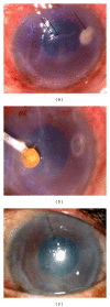
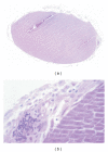
The differential diagnoses for phacolytic glaucoma are acute angle closure glaucoma, open angle glaucoma with uveitis, neovascular glaucoma, and glaucoma secondary to trauma. We report an unusual case where the dislocated cataractous lens firmly adherent to the corneal endothelium evoked a cellular reaction similar to phacolytic glaucoma but clinically appeared like a deep corneal abscess. The 73-year-old lady presented with severe photophobia, pain, and redness in the left eye for two months despite being on antibiotics and antifungals. Anterior chamber wash revealed a cataractous lens buried within the infiltrate, which was removed and sent for histopathological examination. Postoperatively she was treated with topical ofloxacin, homatropine, dorzolamide, timolol, and tapering dose of steroids. Histological confirmation of inflammation, histiocytic response, and giant cells around the lens material confirmed the ongoing phacolytic process. Photophobia, pain, and redness subsided following removal of the lens and surrounding cellular reaction. At her last visit, four months after surgery, she was comfortable.
Figures


References
-
- Brooks AMV, Grant G, Gillies WE. Comparison of specular microscopy and examination of aspirate in phacolytic glaucoma. Ophthalmology. 1990;97(1):85–89. - PubMed
-
- Pradhan D, Hennig A, Kumar J, Foster A. A prospective study of 413 cases of lens-induced glaucoma in Nepal. Indian Journal of Ophthalmology. 2001;49(2):103–107. - PubMed
-
- Allingham RR, Damji K, Freedman S, Moroi S. Sheilds’ Textbook of Glaucoma. 5th edition. chapter 18. Philadelphia, Pa, USA: Lippincott Williams & Wilkins; 2005. Shafranov G.Glaucomas associated with disorders of the Lens; pp. 323–324.
-
- Sridhar MS, Sharma S, Gopinathan U, Rao GN. Anterior chamber tap: diagnostic and therapeutic indications in the management of ocular infections. Cornea. 2002;21(7):718–722. - PubMed
Publication types
LinkOut - more resources
Full Text Sources

