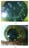
Sands of sahara after LASIK in avellino corneal dystrophy
- PMID: 22606493
- PMCID: PMC3350253
- DOI: 10.1155/2012/413010
Sands of sahara after LASIK in avellino corneal dystrophy
Abstract
We report the case of a patient diagnosed with Avellino corneal dystrophy (ACD) who developed diffuse interstitial keratitis following excimer laser insitu keratomileusis (LASIK). ACD is an autosomal dominant corneal dystrophy characterized by multiple asymmetric stromal opacities that impair vision. Accepted treatments for this condition include corneal transplantation and phototherapeutic keratectomy (PTK). Our patient underwent LASIK at another institution to correct myopia. LASIK and photorefractive keratectomy (PRK) are usually contraindicated in ACD for the high risk of disease recurrence and postoperative complications. The patient came to our attention lamenting blurry vision, decreased visual acuity, and photophobia. Ophthalmologic examination revealed bilateral interstitial keratitis, also known as "sands of Sahara", a seldom-seen complication of LASIK characterized by fine and diffuse granular infiltrates at the surgical flap interface.The risk of developing interstitial keratitis, as in the case presented here, represents another valid reason for avoiding LASIK in patients with ACD.
Figures


Similar articles
-
Comparison of corneal deposits after LASIK and PRK in eyes with granular corneal dystrophy type II.J Refract Surg. 2008 Apr;24(4):392-5. doi: 10.3928/1081597X-20080401-13. J Refract Surg. 2008. PMID: 18500090
-
Avellino corneal dystrophy after LASIK.Ophthalmology. 2004 Mar;111(3):463-8. doi: 10.1016/j.ophtha.2003.06.026. Ophthalmology. 2004. PMID: 15019320
-
Excimer laser in situ keratomileusis and photorefractive keratectomy for correction of high myopia.J Refract Corneal Surg. 1994 Sep-Oct;10(5):498-510. J Refract Corneal Surg. 1994. PMID: 7530099
-
LASIK and surface ablation in corneal dystrophies.Surv Ophthalmol. 2015 Mar-Apr;60(2):115-22. doi: 10.1016/j.survophthal.2014.08.003. Epub 2014 Aug 23. Surv Ophthalmol. 2015. PMID: 25307289 Review.
-
Review of Laser Vision Correction (LASIK, PRK and SMILE) with Simultaneous Accelerated Corneal Crosslinking - Long-term Results.Curr Eye Res. 2019 Nov;44(11):1171-1180. doi: 10.1080/02713683.2019.1656749. Epub 2019 Aug 23. Curr Eye Res. 2019. PMID: 31411927 Review.
Cited by
-
Deterioration of Avellino corneal dystrophy in a Chinese family after LASIK.Int J Ophthalmol. 2021 Jun 18;14(6):795-799. doi: 10.18240/ijo.2021.06.02. eCollection 2021. Int J Ophthalmol. 2021. PMID: 34150532 Free PMC article.
References
-
- Han KE, Kim TI, Chung WS, Choi SI, Kim BY, Kim EK. Clinical findings and treatments of granular corneal dystrophy type 2 (Avellino corneal dystrophy): a review of the literature. Eye and Contact Lens. 2010;36(5):296–299. - PubMed
-
- Park M, Kim DJ, Kim KJ, et al. Genetic associations of common deletion polymorphisms in families with Avellino corneal dystrophy. Biochemical and Biophysical Research Communications. 2009;387(4):688–693. - PubMed
-
- Moon JW, Kim SW, Kim TI, Cristol SM, Chung ES, Kim EK. Homozygous granular corneal dystrophy type II (Avellino corneal dystrophy): natural history and progression after treatment. Cornea. 2007;26(9):1095–1100. - PubMed
-
- Lee ES, Kim EK. Surgical do’s and don’ts of corneal dystrophies. Current Opinion in Ophthalmology. 2003;14(4):186–191. - PubMed
-
- Kim TI, Kim T, Kim SW, Kim EK. Comparison of corneal deposits after LASIK and PRK in eyes with granular corneal dystrophy type II. Journal of Refractive Surgery. 2008;24(4):392–395. - PubMed
Publication types
LinkOut - more resources
Full Text Sources
Molecular Biology Databases

