

Case Reports
doi: 10.1155/2011/687203.
Epub 2011 Sep 22.
Imaging of a case of extramedullary solitary plasmacytoma of the trachea
Affiliations
- PMID: 22606554
- PMCID: PMC3350125
- DOI: 10.1155/2011/687203
Item in Clipboard
Case Reports
Imaging of a case of extramedullary solitary plasmacytoma of the trachea
Case Rep Radiol.
2011.
Abstract
We describe a case of extramedullary tracheal plasmacytoma that was incidentally discovered in a 73-year-old man on a PET scan performed for assessing the extent of colon cancer. CT scan showed the tumor; multiplanar reformation coupled with virtual bronchoscopy allowed proper treatment planning. The tracheal tumor was resected during rigid bronchoscopy. Relevant investigations excluded multiple myeloma. Follow-up CT showed persistent thickening of the tracheal wall, but there has been no recurrence after one-year followup.
Figures

PET scan shows a localized FDG uptake in the middle third of the trachea. (a): PET, (b): CT, (c): PET-CT. (1): axial view, (2): coronal, (3): sagittal.
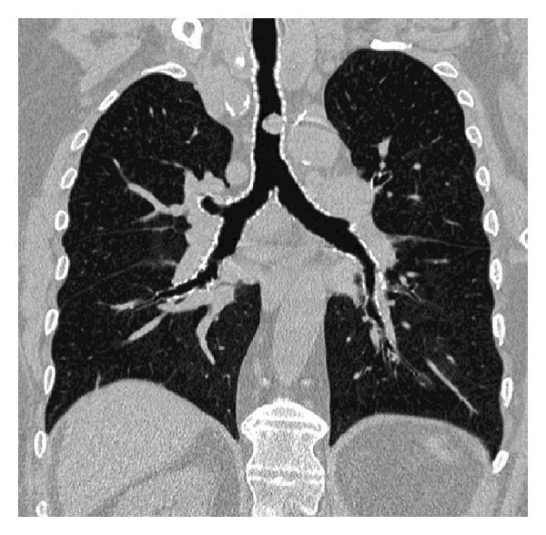
CT scan. CT coronal reformation shows the longitudinal extent of the tumor, its location 35 mm above the carina, and the severity of tracheal narrowing (40%).
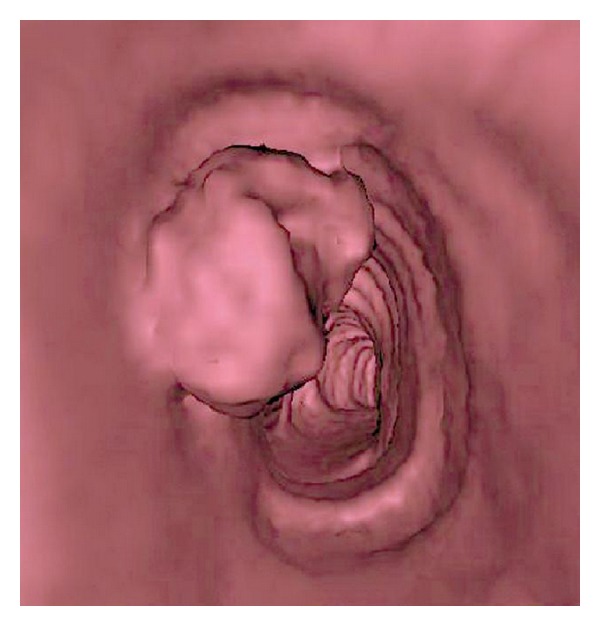
CT scan. Virtual endoscopy demonstrates an endoluminal view of the tracheal tumor.
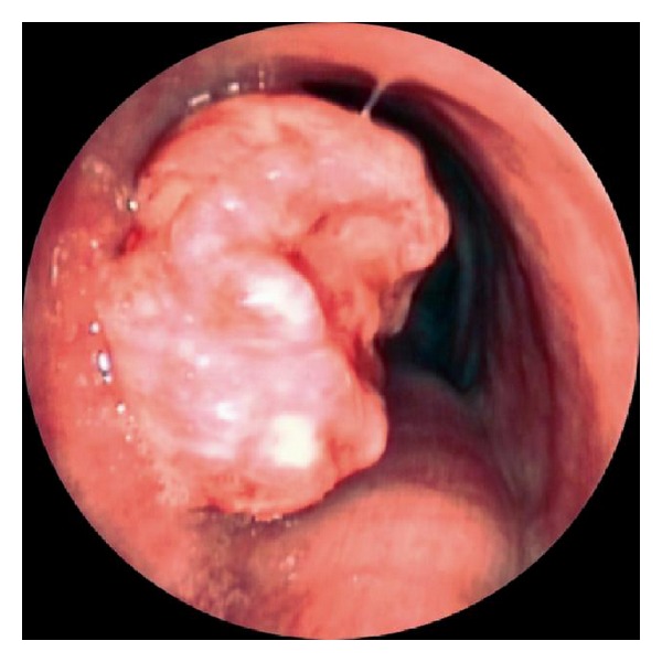
Snapshot by tracheoscopy during the surgery shows an obstructive and fleshy tracheal mass.
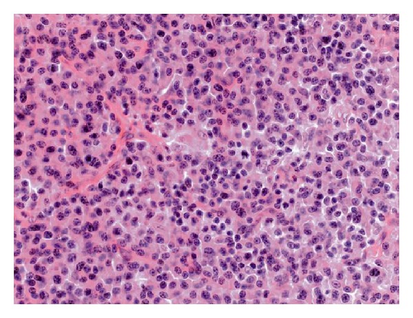
Photomicrograph of surgical specimen. Diffuse infiltrate of neoplastic monoclonal well-differentiated plasma cells is present associated with many deposits of amyloid in the stroma (HES ×200).
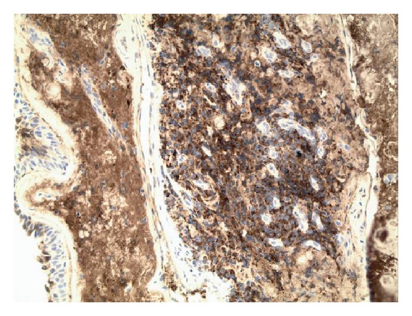
Photomicrograph of surgical specimen. The plasma cells express cytoplasmic immunoglobulin with light chain restriction. They also express CD138, marker characteristically positive for plasma cells (immunohistochemistry, kappa ×200).
References
-
- Dines DE, Lillie JC, Henderson LL, Stickney JM. Solitary plasmacytoma of the trachea. American Review of Respiratory Disease. 1965;92(6):949–951. - PubMed
-
- Rai SP, Kumar R, Bharadwaj R, Panda BN. Solitary tracheal plasmacytoma. The Indian Journal of Chest Diseases & Allied Sciences. 2003;45(4):269–272. - PubMed
-
- Ferretti GR, Bithigoffer C, Righini CA, Arbib F, Lantuejoul S, Jankowski A. Imaging of tumors of the trachea and central bronchi. Thoracic Surgery Clinics. 2010;20(1):31–45. - PubMed
Publication types
LinkOut - more resources
Full Text Sources
Medical
