

Case Reports
doi: 10.1155/2011/481654.
Epub 2011 Sep 6.
Spindle cell hemangioendothelioma of the temporal muscle resected with zygomatic osteotomy: a case report of an unusual intramuscular lesion mimicking sarcoma
Affiliations
- PMID: 22606579
- PMCID: PMC3350060
- DOI: 10.1155/2011/481654
Item in Clipboard
Case Reports
Spindle cell hemangioendothelioma of the temporal muscle resected with zygomatic osteotomy: a case report of an unusual intramuscular lesion mimicking sarcoma
Case Rep Surg.
2011.
Abstract
Spindle cell hemangioendothelioma (SCH) was originally described by Weiss and Enzinger (1986) as a low-grade angiosarcoma resembling both cavernous hemangioma and Kaposi's sarcoma. Recent studies suggest that SCH is a benign neoplasm or reactive lesion accompanying a congenital or acquired vascular malformation. Most SCHs present as one or more nodules affecting the dermis or subcutis of the distal extremities. Few reports describe SCH of the head and neck region; even fewer note intramuscular SCH. Here, we describe a case of SCH involving the temporal muscle mimicking soft tissue sarcoma, who had a successful surgical treatment with a coronal approach and zygomatic osteotomy.
Figures

Preoperative view of the patient.
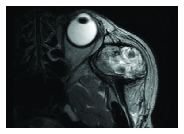
Preoperative MRI on T2-weighted image showing high signal intensity admixed with irregular low signal areas in left temporal fossa.

Preoperative contrast CT showing ill-demarcated low-density mass with enhancement of the adjacent temporal muscle.

Intraoperative view demonstrating intramuscular tumor (arrows) with the assistance of zygomatic osteotomy (asterisks).

Microscopic findings of the spindle cell hemangioendothelioma. (a) Low-power view of the lesion showing cavernous blood vessels filled partly or completely with erythrocytes or thrombus admixed with cellular zones (hematoxylin-eosin, original magnification ×40). (b) High-power view of juxtaposition of the cavernous and cellular areas illustrating blood-filled dilated vessels lined by flattened endothelial cells and spindle-shaped cellular components. There is no evidence of abnormal mitotic activity or nuclear atypia in spindled cells (hematoxylin-eosin, original magnification ×100).

Postoperative view at 24 months followup.
References
-
- Weiss SW, Enzinger FM. Spindle cell hemangioendothelioma. A low-grade angiosarcoma resembling a cavernous hemangioma and Kaposi’s sarcoma. American Journal of Surgical Pathology. 1986;10(8):521–530. - PubMed
-
- Eltorky M, Chesney TM, Sebes J, Hall JC. Spindle cell hemangioendothelioma: report of three cases and review of the literature. Journal of Dermatologic Surgery and Oncology. 1994;20(3):196–202. - PubMed
-
- Terashi H, Itami S, Kurata S, Sonoda T, Takayasu S, Yokoyama S. Spindle cell hemangioendothelioma: report of three cases. Journal of Dermatology. 1991;18(2):104–111. - PubMed
-
- Tosios KI, Gouveris I, Sklavounou A, Koutlas IG. Spindle cell hemangioma (hemangioendothelioma) of the head and neck: case report of an unusual (or underdiagnosed) tumor. Oral Surgery, Oral Medicine, Oral Pathology, Oral Radiology and Endodontology. 2008;105(2):216–221. - PubMed
-
- Isayama T, Iwasaki H, Ogata K, Naito M. Intramuscular spindle cell hemangioendothelioma. Skeletal Radiology. 1999;28(8):477–480. - PubMed
Publication types
LinkOut - more resources
Full Text Sources
Miscellaneous
