. 2012 May 18:9:95.
doi: 10.1186/1742-2094-9-95.

Infection and upregulation of proinflammatory cytokines in human brain vascular pericytes by human cytomegalovirus

Affiliations

Infection and upregulation of proinflammatory cytokines in human brain vascular pericytes by human cytomegalovirus

Donald J Alcendor et al. J Neuroinflammation. .

Abstract

Background: Congenital human cytomegalovirus (HCMV) infections can result in CNS abnormalities in newborn babies including vision loss, mental retardation, motor deficits, seizures, and hearing loss. Brain pericytes play an essential role in the development and function of the blood-brain barrier yet their unique role in HCMV dissemination and neuropathlogy has not been reported.

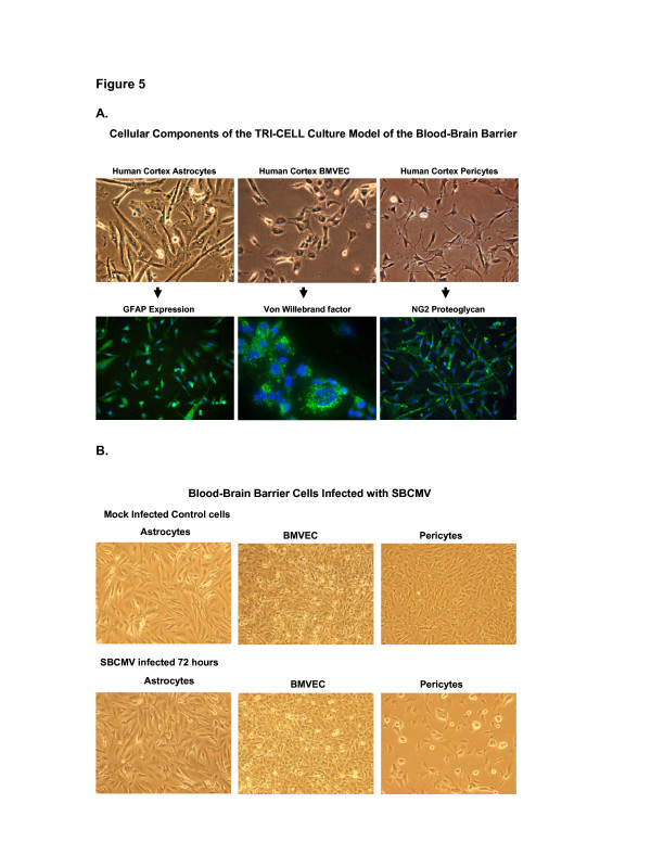
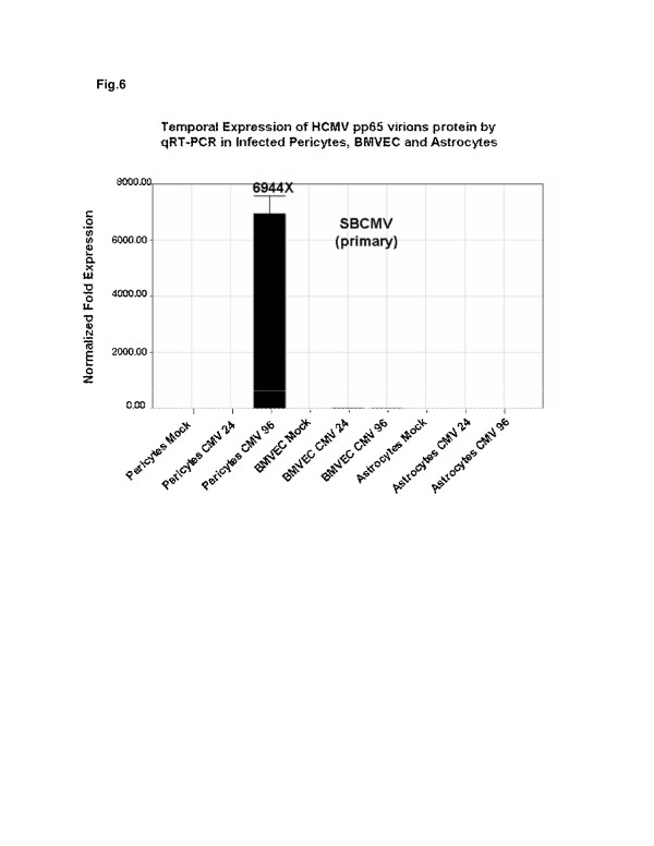
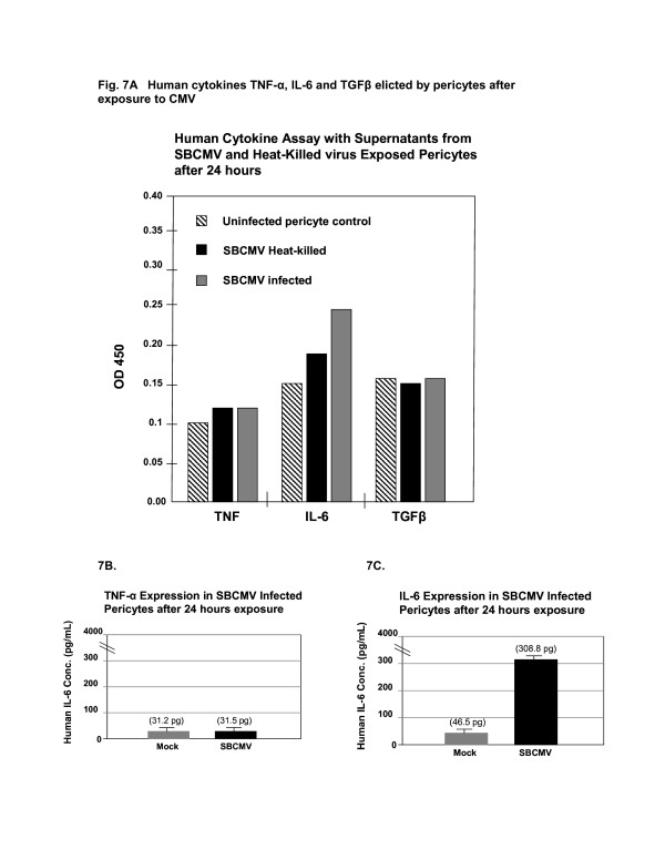
Methods: Primary human brain vascular pericytes were exposed to a primary clinical isolate of HCMV designated 'SBCMV'. Infectivity was analyzed by microscopy, immunofluorescence, Western blot, and qRT-PCR. Microarrays were performed to identify proinflammatory cytokines upregulated after SBCMV exposure, and the results validated by real-time quantitative polymerase chain reaction (qPCR) methodology. In situ cytokine expression of pericytes after exposure to HCMV was examined by ELISA and in vivo evidence of HCMV infection of brain pericytes was shown by dual-labeled immunohistochemistry.

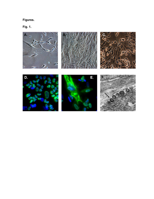
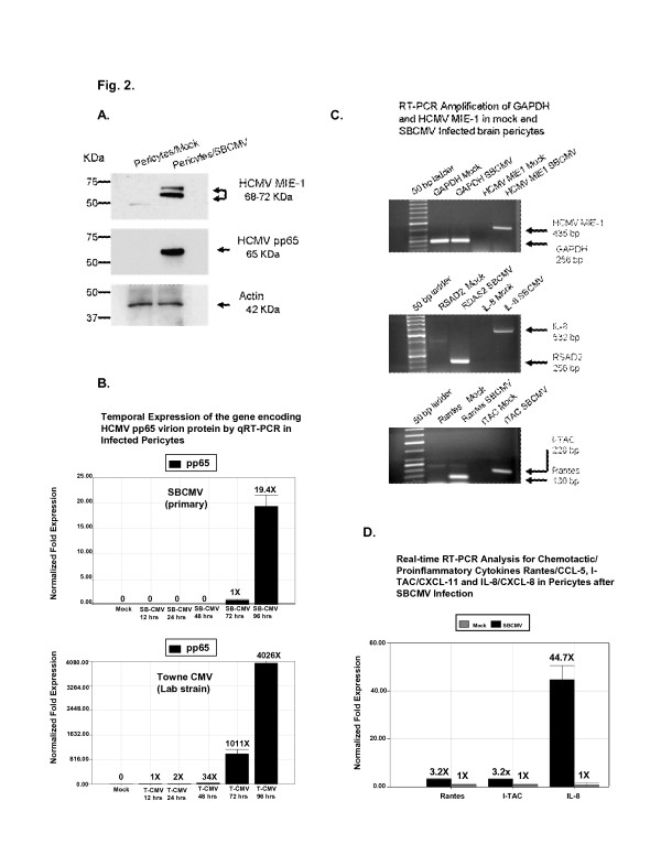
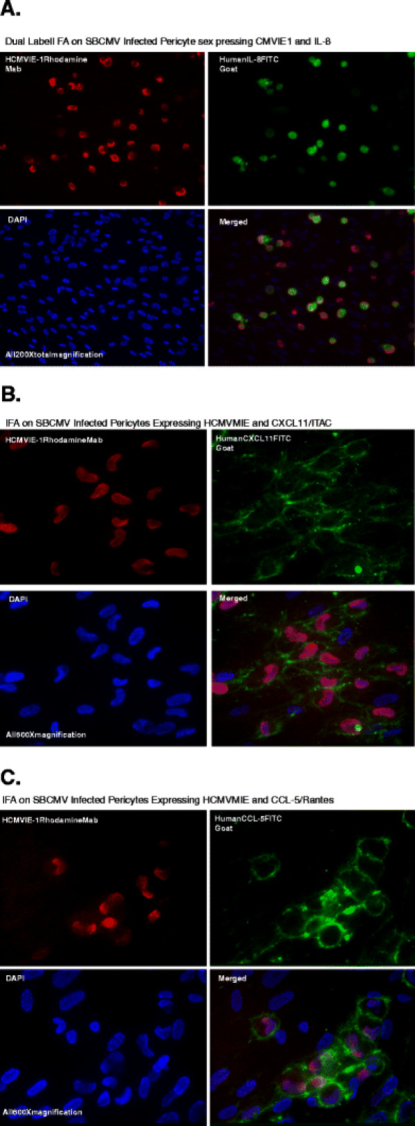
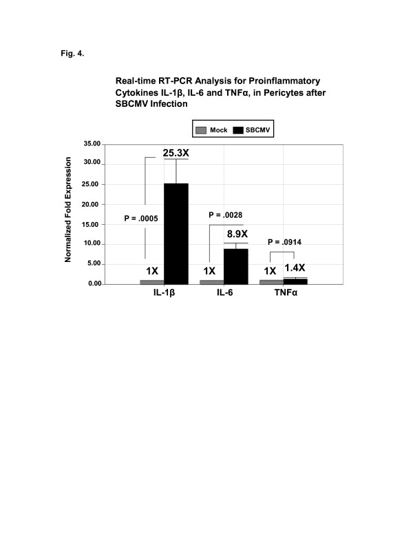
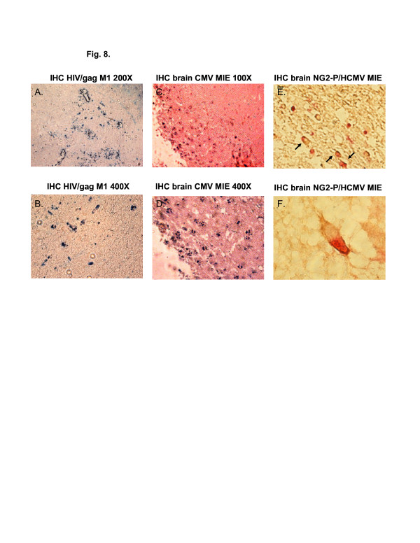
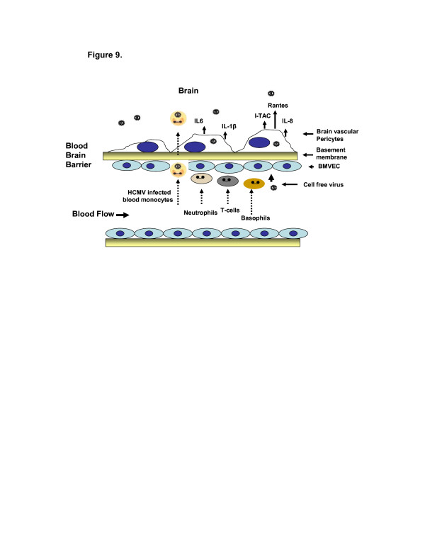
Results: HCMV-infected human brain vascular pericytes as evidenced by several markers. Using a clinical isolate of HCMV (SBCMV), microscopy of infected pericytes showed virion production and typical cytomegalic cytopathology. This finding was confirmed by the expression of major immediate early and late virion proteins and by the presence of HCMV mRNA. Brain pericytes were fully permissive for CMV lytic replication after 72 to 96 hours in culture compared to human astrocytes or human brain microvascular endothelial cells (BMVEC). However, temporal transcriptional expression of pp65 virion protein after SBCMV infection was lower than that seen with the HCMV Towne laboratory strain. Using RT-PCR and dual-labeled immunofluorescence, proinflammatory cytokines CXCL8/IL-8, CXCL11/ITAC, and CCL5/Rantes were upregulated in SBCMV-infected cells, as were tumor necrosis factor-alpha (TNF-alpha), interleukin-1 beta (IL-1beta), and interleukin-6 (IL-6). Pericytes exposed to SBCMV elicited higher levels of IL-6 compared to both mock-infected as well as heat-killed virus controls. A 6.6-fold induction of IL-6 and no induction TNF-alpha was observed in SBCMV-infected cell supernatants at 24 hours postinfection. Using archival brain tissue from a patient coinfected with HCMV and HIV, we also found evidence of HCMV infection of pericytes using dual-label immunohistochemistry, as monitored by NG2 proteoglycan staining.

Conclusion: HCMV lytic infection of primary human brain pericytes suggests that pericytes contribute to both virus dissemination in the CNS as well as neuroinflammation.


Figures

Figure 1
Figure 1
Primary brain vascular pericytes. Phase contrast images of: (A) an uninfected subconfluent monolayer of primary brain vascular pericytes, (B) a confluent monolayer of brain vascular pericytes, and (C) pericytes 72 hours after infection with SBCMV. Immunofluorescence staining of SBCMV-infected pericytes for (D) HCMV MIE protein and (E) pp65 late protein. (F) TEM of SBCMV-infected pericytes showing HCMV virions in the cytoplasm (see arrow). With the exception of the TEM, images were taken on a Nikon TE2000S microscope (200x magnification). HCMV, human cytomegalovirus; MIE, major immediate early protein; SBCMV, primary HCMV isolate from a patient; TEM, transmission electron microscopy.
Figure 2
Figure 2
Protein expression in mock- and SBCMV-infected brain pericytes. (A) Western blot showing expression of HCMV MIE and pp65 virion proteins in pericytes infected with SBCMV for 72 hours. Actin was the loading control. (B) Temporal expression of HCMV pp65 virion protein by real-time PCR in infected pericytes (SBCMV or Towne CMV) at 12, 24, 48, 72, and 96 hours postinfection. (C) Semi-quantitative RT-PCR for HCMV MIE, GAPDH, CXCL8/IL-8, RSAD2, CXCL11/I-TAC and CCL5/Rantes (72-hour exposure). GAPDH was the loading control. (D) Real time RT-PCR analysis for CCL5/Rantes, CXCL11/I-TAC, and CXCL8/IL-8 in pericytes 72 hours after SBCMV infection. This experiment was replicated three times, and amplifications were performed in triplicate and normalized to GAPDH. CCL5/Rantes, chemokine (C-C motif) ligand 5/Rantes; CXCL8/IL-8, blood–brain barrier; CXCL11/I-TAC, chemokine (C-X-C motif) ligand 11/I-TAC; GAPDH, gyceralaldehyde phosphate dehydrogenase; HCVM, human cytomegalovirus; MIE, major immediate early protein: pp65, human cytomegalovirus phosphorylated envelop protein expressed at late times during virus replication; RSAD2, radical S-adenosyl methionine domain-containing protein 2; RT-PCR, reverse transcription polymerase chain reaction; SBCMV, primary HCMV isolate from a patient.
Figure 3
Figure 3
Dual-labeled immunofluorescent staining of SBCMV-infected pericytes showing colocalization (merged images) of proteins. (A) Infected pericytes expressing MIE proteins and CXCL8/IL-8. (B) Infected pericytes expressing MIE proteins and CXCL11/I-TAC. (C) Infected pericytes expressing MIE proteins and CCL5/Rantes. CCL5/Rantes, chemokine (C-C motif) ligand 5/Rantes; CXCL8/IL-8, blood–brain barrier; CXCL11/I-TAC, chemokine (C-X-C motif) ligand 11/I-TAC; MIE. major immediate early protein; SBCMV, primary HCMV isolate from a patient.
Figure 4
Figure 4
Real time RT-PCR analysis for proinflammatory cytokines. Cytokine IL-1 beta, IL-6 and TNF-alpha expression in pericytes 72 hours after SBCMV infection. This experiment was replicated three times, and amplifications were performed in triplicate and normalized to GAPDH. GAPDH, gyceralaldehyde phosphate dehydrogenase; RT-PCR, reverse transcription polymerase chain reaction; SBCMV, primary HCMV isolate from a patient.
Figure 5
Figure 5
CMV infection of blood–brain barrier cellular components. Human brain cortical astrocytes, BMVEC and pericytes representing the major cellular components of the blood–brain barrier were infected with SBCMV. Cells cultivated to 50% confluence were stained with representative markers. (A) Astrocytes were stained for glial fibillary acidic protein (GFAP), BMVEC for von Willebrand factor (vWF) and brain pericytes with NG2 proteoglycan antibodies. (B) All three cell types were infected with SBCMV for 72 hours. BMVEC, brain microvascular endothelial cells; GFAP, glial fibrillary acidic protein; NG2, neuron-glial antigen 2; SBCMV, primary HCMV isolate from a patient; vWF, von Willebrand factor.
Figure 6
Figure 6
Expression of SBCMV pp65 virion protein mRNA by pericytes, BMVEC and astrocytes. Real-time PCR analysis of pp65 late virion protein mRNA levels in SBCMV-infected pericytes, BMVEC and astrocytes. cDNA was prepared from total RNA isolated at 24 and 96 hours postinfection along with RNA from mock-infected control cells. Black bars indicate fold transcription for SBCMV-infected pericytes, BMVEC and astrocytes. This experiment was replicated three times and normalized to GAPDH. BMVEC, brain microvascular endothelial cells; GAPDH,gyceralaldehyde phosphate dehydrogenase; pp65, human cytomegalovirus phosphorylated envelop protein expressed at late times during virus replication; PCR, polymerase chain reaction; SBCMV, primary HCMV isolate from a patient.
Figure 7
Figure 7
Human cytokine ELISA assay. Analysis of human cytokines after SBCMV infection of brain vascular pericytes. A human cytokine ELISA assay was performed on supernatants from SBCMV-infected pericytes, SBCMV heat-killed pericytes and mock-infected cells. Shown are the relative amounts of TNF-alpha, IL-6 and TGF-beta in cell culture supernatants elicited 24 hours postexposure (A). Quantitative measurements IL-6 and TNF-alpha secreted from pericytes in infected cell supernatants 24 hours postinfection was based on optical density utilizing a standard curve for human IL-6 (B) and human TNF-alpha (C). Analysis was performed in triplicate and values were expressed in picograms/milliliter. IL, interleukin; SBCMV, primary HCMV isolate from a patient; TGF-beta, tumor growth factor-beta; TNF-alpha, tumor necrosis factor-alpha.
Figure 8
Figure 8
Immunohistochemical staining of HIV and CMV coinfected brain tissue. IHC staining of archival human hippocampus brain tissue coinfected with HCMV from an HIV-infected patient. (A/B) Brain tissue IHC-stained for HIV Gag) using True Blue (Kirkegaard & Perry Laboratories, Gaithersburg, MD, USA) as a substrate for immunoperoxidase. Photographs in A and B were taken at 200x and 400x magnification, respectively. (D/C) IHC-stained brain tissue for the HCMV MIE gene protein using True Blue as an immunoperoxidase substrate. Photographs were at 200x and 400x magnification. (E) Brain tissue IHC dual-stained for the pericyte marker NG2 proteogylcan using 3,3-diaminobenzidine (DAB) staining cells brown and CMV MIE gene protein using Vector Red (Vector Laboratories, Burlingame, CA, USA) as a substrate for alkaline phosphatase (dual-stained indicated by arrows). This photograph was taken at 200x magnification. (F) Brain tissue dual-stained by IHC for NG2 proteoglycan and HCMV MIE gene protein showing a cell with a brown-colored cytoplasm and a red nucleus. This image was taken at 600x magnification. All images were taken with a Nikon TE2000S microscope fitted with a CCD camera (Nikon, Tokyo, Japan). CMV, cytomegalovirus; DAB, 3,3-diaminobenzidine; IHC, immunohistochemistry; HCMV, human cytomegalovirus; MIE, major immediate early protein; NG2, neuron-glial antigen 2.
Figure 9
Figure 9
Model of HCMV dissemination across the blood–brain barrier including pericytes and a CMV-elicited proinflammatory cytokine cascade. A working model of HCMV dissemination across the blood–brain barrier involving pericytes and a proinflammatory cytokine cascade. (H)CMV, (human) cytomegalovirus.

Similar articles

Cited by

References

    1. Boppana SB, Fowler KB, Pass RF, Rivera LB, Bradford RD, Lakeman FD, Britt WJ. Congenital cytomegalovirus infection: association between virus burden in infancy and hearing loss. J Pediatr. 2005;146:817–823. doi: 10.1016/j.jpeds.2005.01.059. - DOI - PubMed
    1. Colugnati FA, Staras SA, Dollard SC, Cannon MJ. Incidence of cytomegalovirus infection among the general population and pregnant women in the United States. BMC Infect Dis. 2007;7:71. doi: 10.1186/1471-2334-7-71. - DOI - PMC - PubMed
    1. Sessions CF, Taeusch HW. In: Viral infections of the newborn. In Diseases of the newborn. Taeusch HW, Ballard RA, Avery ME, editor. W. B. Saunders, Philadelphia; 1991. pp. 331–349.
    1. Stagno S, Pass RF, Dworsky ME, Henderson RE, Moore EG, Walton PD, Alford CA. Congenital cytomegalovirus infection: the relative importance of primary and recurrent maternal infection. N Engl J Med. 1982;306:945–949. doi: 10.1056/NEJM198204223061601. - DOI - PubMed
    1. Demmler GJ. Congenital cytomegalovirus infection and disease. Adv Pediatr Infect Dis. 1996;11:135–162. - PubMed

Publication types

MeSH terms