

Microbeam radiation therapy alters vascular architecture and tumor oxygenation and is enhanced by a galectin-1 targeted anti-angiogenic peptide
- PMID: 22607585
- PMCID: PMC3391740
- DOI: 10.1667/rr2784.1
Microbeam radiation therapy alters vascular architecture and tumor oxygenation and is enhanced by a galectin-1 targeted anti-angiogenic peptide
Abstract
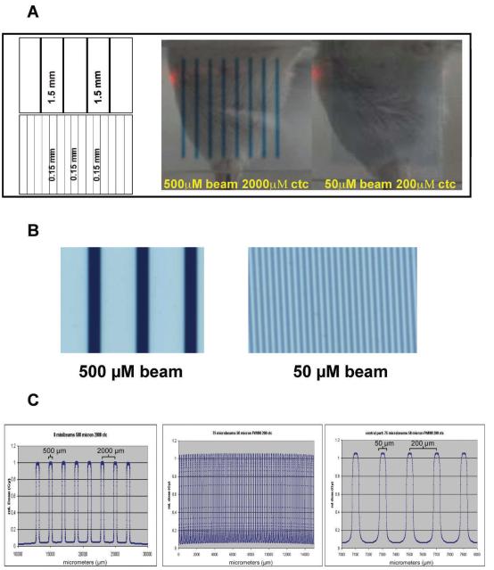
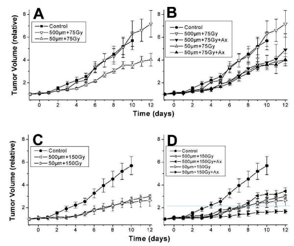
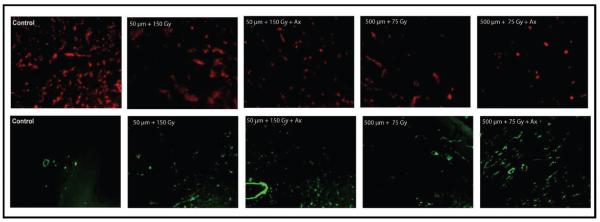
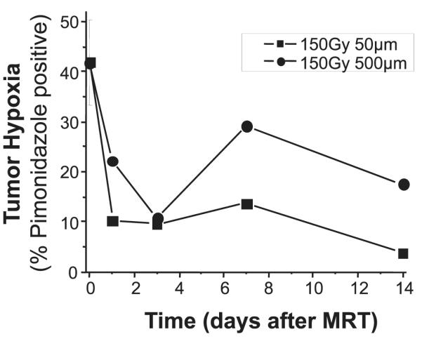
In this study, we sought to determine the therapeutic potential of variably sized (50 μm or 500 μm wide, 14 mm tall) parallel microbeam radiation therapy (MRT) alone and in combination with a novel anti-angiogenic peptide, anginex, in mouse mammary carcinomas (4T1)--a moderately hypoxic and radioresistant tumor with propensity to metastasize. The fraction of total tumor volume that was directly irradiated was approximately 25% in each case, but the distance between segments irradiated by the planar microbeams (width of valley dose region) varied by an order of magnitude from 150-1500 μm corresponding to 200 μm and 2000 μm center-to-center inter-microbeam distances, respectively. We found that MRT administered in 50 μm beams at 150 Gy was most effective in delaying tumor growth. Furthermore, tumor growth delay induced by 50 μm beams at 150 Gy was virtually indistinguishable from the 500 μm beams at 150 Gy. Fifty-micrometer beams at the lower peak dose of 75 Gy induced growth delay intermediate between 150 Gy and untreated tumors, while 500 μm beams at 75 Gy were unable to alter tumor growth compared to untreated tumors. However, the addition of anginex treatment increased the relative tumor growth delay after 500 μm beams at 75 Gy most substantially out of the conditions tested. Anginex treatment of animals whose tumors received the 50 μm beams at 150 Gy also led to an improvement in growth delay from that induced by the comparable MRT alone. Immunohistochemical staining for CD31 (endothelial cells) and αSMA (smooth muscle pericyte-associated blood vessels as a measure of vessel normalization) indicated that vessel density was significantly decreased in all irradiated groups and pericyte staining was significantly increased in the irradiated groups on day 14 after irradiation. The addition of anginex treatment further decreased the mean vascular density in all combination treatment groups and further increased the amount of pericyte staining in these tumors. Finally, evidence of tumor hypoxia was found to decrease in tumors analyzed at 1-14 days after MRT in the groups receiving 150 Gy peak dose, but not 75 Gy peak dose. Our results suggest that tumor vascular damage induced by MRT at these potentially clinically acceptable peak entrance doses may provoke vascular normalization and may be exploited to improve tumor control using agents targeting angiogenesis.
Figures
Similar articles
-
Anginex synergizes with radiation therapy to inhibit tumor growth by radiosensitizing endothelial cells.Int J Cancer. 2005 Jun 10;115(2):312-9. doi: 10.1002/ijc.20850. Int J Cancer. 2005. PMID: 15688384
-
Antiangiogenesis therapy using a novel angiogenesis inhibitor, anginex, following radiation causes tumor growth delay.Int J Clin Oncol. 2007 Feb;12(1):42-7. doi: 10.1007/s10147-006-0625-y. Epub 2007 Feb 25. Int J Clin Oncol. 2007. PMID: 17380440
-
Effects of pulsed, spatially fractionated, microscopic synchrotron X-ray beams on normal and tumoral brain tissue.Mutat Res. 2010 Apr-Jun;704(1-3):160-6. doi: 10.1016/j.mrrev.2009.12.003. Epub 2009 Dec 23. Mutat Res. 2010. PMID: 20034592 Review.
-
Synchrotron microbeam radiation therapy for rat brain tumor palliation-influence of the microbeam width at constant valley dose.Phys Med Biol. 2009 Nov 7;54(21):6711-24. doi: 10.1088/0031-9155/54/21/017. Epub 2009 Oct 20. Phys Med Biol. 2009. PMID: 19841517
-
Advances and prospects of anginex as a promising anti-angiogenesis and anti-tumor agent.Peptides. 2012 Dec;38(2):457-62. doi: 10.1016/j.peptides.2012.09.007. Epub 2012 Sep 14. Peptides. 2012. PMID: 22985857 Review.
Cited by
-
Reduced side effects by proton microchannel radiotherapy: study in a human skin model.Radiat Environ Biophys. 2013 Mar;52(1):123-33. doi: 10.1007/s00411-012-0450-9. Epub 2012 Dec 28. Radiat Environ Biophys. 2013. PMID: 23271171
-
Longitudinally Heterogeneous Tumor Dose Optimizes Proton Broadbeam, Interlaced Minibeam, and FLASH Therapy.Cancers (Basel). 2022 Oct 21;14(20):5162. doi: 10.3390/cancers14205162. Cancers (Basel). 2022. PMID: 36291946 Free PMC article.
-
Neuro-Oncologic Veterinary Trial for the Clinical Transfer of Microbeam Radiation Therapy: Acute to Subacute Radiotolerance after Brain Tumor Irradiation in Pet Dogs.Cancers (Basel). 2024 Jul 29;16(15):2701. doi: 10.3390/cancers16152701. Cancers (Basel). 2024. PMID: 39123429 Free PMC article.
-
Galectins as Molecular Targets for Therapeutic Intervention.Int J Mol Sci. 2018 Mar 19;19(3):905. doi: 10.3390/ijms19030905. Int J Mol Sci. 2018. PMID: 29562695 Free PMC article. Review.
-
Superior Anti-Tumor Response After Microbeam and Minibeam Radiation Therapy in a Lung Cancer Mouse Model.Cancers (Basel). 2025 Jan 1;17(1):114. doi: 10.3390/cancers17010114. Cancers (Basel). 2025. PMID: 39796741 Free PMC article.
References
-
- Dilmanian FA, Morris GM, Zhong N, Bacarian T, Hainfeld JF, Kalef-Ezra J, et al. Murine EMT-6 carcinoma: high therapeutic efficacy of microbeam radiation therapy. Radiat Res. 2003;159(5):632–41. - PubMed
-
- Smilowitz HM, Blattmann H, Brauer-Krisch E, Bravin A, Di Michiel M, Gebbers JO, et al. Synergy of gene-mediated immunoprophylaxis and microbeam radiation therapy for advanced intracerebral rat 9L gliosarcomas. J Neuro-Oncology. 2006;78(2):135–43. - PubMed
-
- Slatkin DN, Spanne P, Dilmanian FA, Sandborg M. Microbeam radiation therapy. Med Phys. 1992;19(6):1395–400. - PubMed
Publication types
MeSH terms
Substances
Grants and funding
LinkOut - more resources
Full Text Sources
Other Literature Sources
Medical
Research Materials
