Meta-Analysis
. 2012 May 18:12:69.
doi: 10.1186/1471-2148-12-69.

Demasculinization of the Anopheles gambiae X chromosome

Affiliations
Meta-Analysis

Demasculinization of the Anopheles gambiae X chromosome

Kalle Magnusson et al. BMC Evol Biol. .

Abstract

Background: In a number of organisms sex-biased genes are non-randomly distributed between autosomes and the shared sex chromosome X (or Z). Studies on Anopheles gambiae have produced conflicting results regarding the underrepresentation of male-biased genes on the X chromosome and it is unclear to what extent sexual antagonism, dosage compensation or X-inactivation in the male germline, the evolutionary forces that have been suggested to affect the chromosomal distribution of sex-biased genes, are operational in Anopheles.

Results: We performed a meta-analysis of sex-biased gene expression in Anopheles gambiae which provides evidence for a general underrepresentation of male-biased genes on the X-chromosome that increased in significance with the observed degree of sex-bias. A phylogenomic comparison between Drosophila melanogaster, Aedes aegypti and Culex quinquefasciatus also indicates that the Anopheles X chromosome strongly disfavours the evolutionary conservation of male-biased expression and that novel male-biased genes are more likely to arise on autosomes. Finally, we demonstrate experimentally that transgenes situated on the Anopheles gambiae X chromosome are transcriptionally silenced in the male germline.

Conclusion: The data presented here support the hypothesis that the observed demasculinization of the Anopheles X chromosome is driven by X-chromosome inactivation in the male germline and by sexual antagonism. The demasculinization appears to be the consequence of a loss of male-biased expression, rather than a failure in the establishment or the extinction of male-biased genes.


Figures
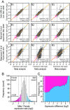
Figure 1
Figure 1
Meta-analysis of genome-wide sex-biased expression in adultA. gambiae.A) Meta analysis of male to female expression ratios and correlation with original expression data from published studies used for metadata computation. R1, R2 and R3 correspond to the biological replicates presented in each study. Each dot represents a gene, with genes presenting 2-fold increased expression in male A. gambiae when compared to females for both metadata and original reaction shown in blue, while genes presenting a 2-fold decrease for the same conditions are shown in pink. B) Histogram presenting the frequency of genes that were identified by the meta analysis for varying male to female expression ratios. C) Proportion of male- (blue) and female-biased genes (pink) and their expression difference in the meta analysis dataset.
Figure 2
Figure 2
Chromosomal linkage of sex-biased genes in adultA. gambiae.A) Percentages of genes with increased expression in adult male (blue) or female (pink) A. gambiae mosquitoes are shown for the chromosomes X, 2 and 3 at increasing expression ratios. Significant differences in the number of genes with increased expression found in a particular chromosome were evaluated by hypergeometric distribution using as reference the overall number of genes linked to the same chromosome in the meta-analysis dataset (orange). B) Percentages of significantly regulated male (blue), female (pink) and non-biased genes (grey), as identified by RankProduct analysis, in the overall dataset, the X chromsome and the autosomes 2 and 3. Significant differences in the number of male and female biased genes observed were evaluated by hypergeometric distribution against the observed number for the same class in the overall dataset (P = <0.01:asterisk). C) Average chromosome-wide expression intensity per chromosome presented for male-biased genes and female-biased genes. Bar graphs presenting the average expression intensity for all genes in a particular chromosome (black line), + − 25th-75th quartile (box) and range of chromosome-wide hybridization intensities (whiskers) are shown.
Figure 3
Figure 3
Reporter gene expression of autosomal and X-linked transgenes inA. gambiae. (A) Transmission and fluorescence (upper panels show RFP, lower panels show GFP) microphotographs of larvae heads and dissected testes taken from β2-eGFP 2 L, β2-eGFP-3R and β2-eGFP X mosquito lines. (B) Quantitative PCR analysis of the transcription activity of the neuronal 3xP3 and the testis specific β2-tubulin promoters. The bars show the ratios of quantitative RT-PCR signals detected in mosquito lines carrying autosomal integrations (β2-eGFP 2 L and β2-eGFP 3R) versus the X linked integration (β2-eGFP X) for the eGFP (green) and the DsRed (red) marker genes. (C) Expression activity of the α-tubulin promoter. Fluorescent microphotographs of six transgenic lines carrying single insertions of the α-tubulin-eGFP construct into either the X chromosome or one of the autosomes. The sites of chromosomal insertions are indicated on the top of the microphotographs. Red arrows in the photographs of the lower panel indicate testis.
Figure 4
Figure 4
Evolution of X-linked and autosomal sex-biased genes. Percentages of male- (blue) and female-biased (pink) genes found to be unique when comparing the genomes of A. gambiae (An) with A. aegypti (Ae), C. quinquefasciatus (Cu) and D. melanogaster (Dm) are presented for X-linked (upper panels) and autosomal genes (lower panels). Statistically significant differences were evaluated by hypergeometric distribution using as reference the fraction of unique genes found in all X-linked or autosomal genes studied (P < 0.05, one asterisk; P < 0.01, two asterisks).
Figure 5
Figure 5
Conservation of sex-biased expression of X-linked and autosomal orthologs inA. gambiae andD. melanogaster. The transcription profile of male-, female and non sex-biased A. gambiae genes was compared to their one-to-one D. melanogaster orthologs. The bars show the proportion of A. gambiae sex biased genes with a D. melanogaster ortholog showing either a conserved male (blue), female (pink) or a reversed (light gray) transcription pattern. The proportion of A. gambiae non sex-biased genes with sex-biased D. melanogaster orthologs is also shown (dark grey). Shown is the percentage of such genes according to their location in both A. gambiae (Ag) and D. melanogaster (Dm) on either the autosomes (Auto) or the X chromosome (X).

Similar articles

Cited by

References

    1. Prince EG, Kirkland D, Demuth JP. Hyperexpression of the X chromosome in both sexes results in extensive female bias of X-linked genes in the flour beetle. Genome Biol Evol. 2010;2:336–346. doi: 10.1093/gbe/evq024. - DOI - PMC - PubMed
    1. Ranz JM, Castillo-Davis CI, Meiklejohn CD, Hartl DL. Sex-dependent gene expression and evolution of the Drosophila transcriptome. Science. 2003;300(5626):1742–1745. doi: 10.1126/science.1085881. - DOI - PubMed
    1. Parisi M, Nuttall R, Naiman D, Bouffard G, Malley J, Andrews J, Eastman S, Oliver B. Paucity of genes on the Drosophila X chromosome showing male-biased expression. Science. 2003;299(5607):697–700. doi: 10.1126/science.1079190. - DOI - PMC - PubMed
    1. Reinke V, Smith HE, Nance J, Wang J, Van Doren C, Begley R, Jones SJ, Davis EB, Scherer S, Ward S. et al.A global profile of germline gene expression in C. elegans. Mol Cell. 2000;6(3):605–616. doi: 10.1016/S1097-2765(00)00059-9. - DOI - PubMed
    1. Storchova R, Divina P. Nonrandom representation of sex-biased genes on chicken Z chromosome. J Mol Evol. 2006;63(5):676–681. doi: 10.1007/s00239-006-0022-1. - DOI - PubMed

Publication types