. 2012 Jun 15;22(12):4038-43.
doi: 10.1016/j.bmcl.2012.04.082. Epub 2012 Apr 26.

5-Cyano-6-oxo-1,6-dihydro-pyrimidines as potent antagonists targeting exchange proteins directly activated by cAMP

Affiliations

5-Cyano-6-oxo-1,6-dihydro-pyrimidines as potent antagonists targeting exchange proteins directly activated by cAMP

Haijun Chen et al. Bioorg Med Chem Lett. .

Abstract

Exchange proteins directly activated by cAMP (Epac) are a family of guanine nucleotide exchange factors that regulate a wide variety of intracellular processes in response to second messenger cAMP. To explore the structural determinants for Epac antagonist properties of high throughput screening (HTS) hit ESI-08, pyrimidine 1, a series of 5-cyano-6-oxo-1,6-dihydro-pyrimidine analogues have been synthesized and evaluated for their activities for Epac inhibition. Structure-activity relationship (SAR) analysis led to the identification of three more potent Epac antagonists (6b, 6g, and 6h). These inhibitors may serve as valuable pharmacological probes for further elucidation of the physiological functions and mechanisms of Epac regulation. Our SAR results and molecular docking studies have also revealed that further optimization of the moieties at the C-6 position of pyrimidine scaffold may allow us to discover more potent Epac-specific antagonists.


Figures

Figure 1
Figure 1
The structures of cAMP, Epac agonists: 2'-O-Me-cAMP analogs 007 and 007-AM, Epac antagonist: HTS hit 1 (ESI-08).
Figure 2
Figure 2. Relative potency of Epac2 antagonists
Dose-dependent competition of Epac2 antagonists with 8-NBD-cAMP in binding to Epac2: closed circles, 6g (HJC0198); closed triangles up, 6h (HJC0197); closed diamonds, 1 (ESI-08); open triangles down, 6a (HJC0167); closed squares, cAMP.
Figure 3
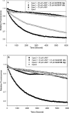
Figure 3. Specificity of Epac antagonists 6g and 6h
(A) cAMP-mediated Epac1 GEF activity measured in the presence or absence of Epac antagonists: open squares, Epac1 alone; closed squares: Epac1 in the presence of 25 μM cAMP; open circles, Epac1 with 25 μM cAMP and 25 μM 6g; closed circles, Epac1 with 25 μM cAMP and 25 ȝM 6h. (B) cAMP-mediated Epac2 GEF activity measured in the presence or absence of Epac antagonists: open squares, Epac2 alone; closed squares: Epac2 in the presence of 25 μM cAMP; open circles, Epac2 with 25 μM cAMP and 25 μM 6g; closed circles, Epac2 with 25 μM cAMP and 25 μM 6h. Similar results were obtained from two independent experiments.
Figure 4
Figure 4. Effects of Epac antagonists ESI-08, 6g (HJC0198) and 6h (HJC0197) on type I and II PKA activities
Relative Type I (filled bars) and II (open bars) PKA holoenzyme activities in the presence of 100 μM cAMP plus vehicle control, 25 μM H89 or 25 μM ESI-08 or 25 μM 6g (HJC0198) or 25 μM 6h (HJC0197). Data are presented in the format of means and standard deviations (n = 3).
Figure 5
Figure 5. Effects of Epac antagonists on Epac-mediated Akt/PKB phosphorylation in HEK293/Epac cells
HEK293/Epac1 and HEK293/Epac2 cells with or without pretreatment of 10 μM Epac antagonists (HJC0197 and HJC0198, respectively) were stimulated with 10 μM 007-AM. Cell lysates were subjected to Western blot analyses using anti-phospho-Ser473-specific (PKB-P473) and anti-phospho-Thr308-specific (PKB-P308) PKB antibodies.
Figure 6
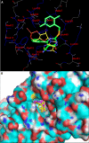
Figure 6. Predicted binding mode and molecular docking of compound 6h into the cAMP binding domain (CBD) of Epac2 protein
(A) Nitrogen, oxygen, sulfur and phosphorus are shown in blue, red, orange and pale red, respectively. 6h is shown in big sticks and in green color. cAMP is shown in small sticks and in yellow color. Protein residues likely to be involved in polar and hydrophobic interactions are shown in sticks. Hydrogen bonds are indicated by dashed lines. (B) 6h is shown in yellow color while cAMP is shown in pink color.
Scheme 1
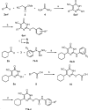
Scheme 1
aReagents and conditions: (a) piperidine, EtOH, reflux; 5a R1 = cyclohexyl, 51%; 5b R1 = isopropyl, 55%; 5c R1 = cyclopropyl, 18%; 5d R1 = cyclopentyl, 62%; 5e R1 = 1-methyl-piperidin-4-yl, 69%; 5f R1 = 1-Boc-piperidin-4-yl, 59%; 8a R3 = H, 48%; 8b R3 = 4-Cl, 49%; (b) various benzyl halides, K2CO3, acetone, rt; 6a 93%, 6b 87%; 6c 91%; 6d 94%; 6e 95%; 6f 75%; 6g 80%; 6h 92%; 6i 65%; 6j 77%; (c) TFA, DCM, 0 °C, 92%; (d) EDCI, DIPEA, various anilines, DCM, rt; 11a 91%; 11b 80%; 11c 82%.

Similar articles

Cited by

References

    1. Beavo JA, Brunton LL. Nat. Rev. Mol. Cell Biol. 2002;3:710. - PubMed
    1. Houslay MD, Kolch W. Mol. Pharmacol. 2000;58:659. - PubMed
    1. Zaccolo M, Movsesian MA. Circ. Res. 2007;100:1569. - PubMed
    1. Bourne HR, Lichtenstein LM, Melmon KL, Henney CS, Weinstein Y, Shearer GM. Science. 1974;184:19. - PubMed
    1. Follin-Arbelet V, Hofgaard PO, Hauglin H, Naderi S, Sundan A, Blomhoff R, Bogen B, Blomhoff HK. BMC Cancer. 2011;11:301. - PMC - PubMed

Publication types

MeSH terms