. 2012 May 18;10(1):33.
doi: 10.1186/1477-5956-10-33.

Vigilin interacts with signal peptide peptidase

Affiliations

Vigilin interacts with signal peptide peptidase

Stephen Hsueh-Jeng Lu et al. Proteome Sci. .

Abstract

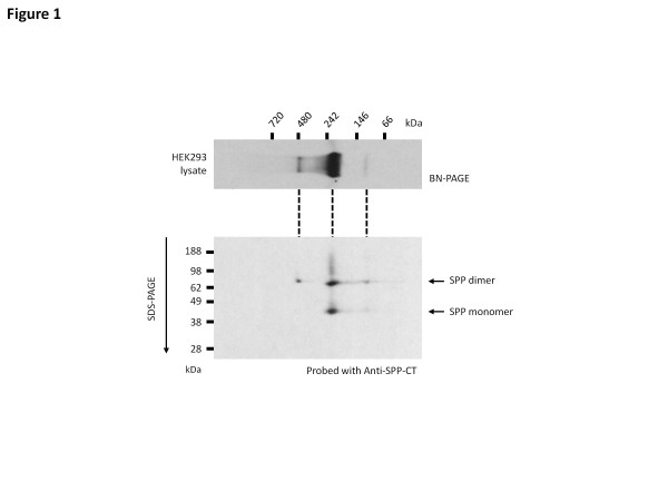
Background: Signal peptide peptidase (SPP), a member of the presenilin-like intra-membrane cleaving aspartyl protease family, migrates on Blue Native (BN) gels as 100 kDa, 200 kDa and 450 kDa species. SPP has recently been implicated in other non-proteolytic functions such as retro-translocation of MHC Class I molecules and binding of misfolded proteins in the endoplasmic reticulum (ER). These high molecular weight SPP complexes might contain additional proteins that regulate the proteolytic activity of SPP or support its non-catalytic functions.

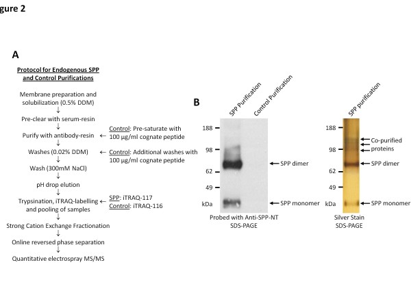
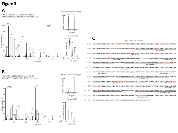
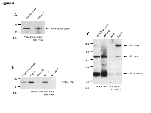
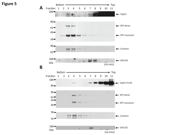
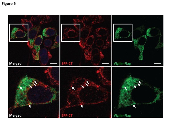
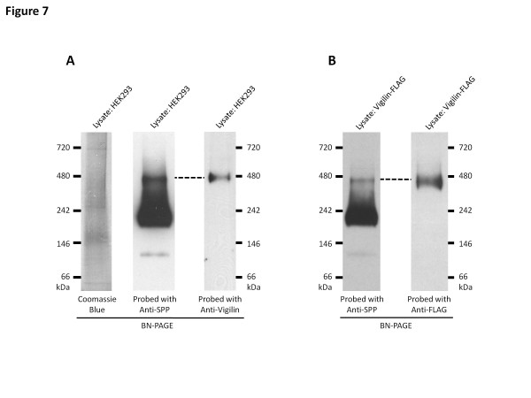
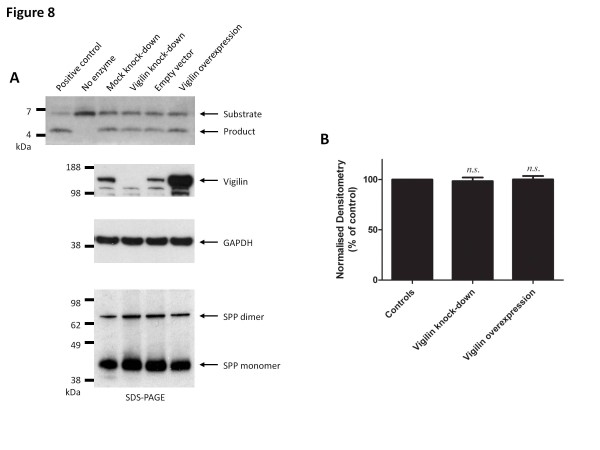
Results: In this study, an unbiased iTRAQ-labeling mass spectrometry approach was used to identify SPP-interacting proteins. We found that vigilin, a ubiquitous multi-KH domain containing cytoplasmic protein involved in RNA binding and protein translation control, selectively enriched with SPP. Vigilin interacted with SPP and both proteins co-localized in restricted intracellular domains near the ER, biochemically co-fractionated and were part of the same 450 kDa complex on BN gels. However, vigilin does not alter the protease activity of SPP, suggesting that the SPP-vigilin interaction might be involved in the non-proteolytic functions of SPP.

Conclusions: We have identified and validated vigilin as a novel interacting partner of SPP that could play an important role in the non-proteolytic functions of SPP. This data adds further weight to the idea that intramembrane-cleaving aspartyl proteases, such as presenilin and SPPs, could have other functions besides the proteolysis of short membrane stubs.


Figures

Figure 1
Figure 1
SPP exists in higher molecular weight complexes.Top panel, HEK293 lysate solubilized in 0.5% DDM and resolved on 16% Bis-Tris Blue Native-PAGE gels, revealing SPP-containing complexes at around 450 kDa, 200 kDa and 100 kDa. Lower panel, lysates resolved on the BN-PAGE were then resolved on the second dimension/SDS-PAGE. SDS-stable SPP dimer can be found in all three high molecular weight bands suggesting that the 100 kDa band observed in the first dimension is a SDS-stable SPP dimer.
Figure 2
Figure 2
Purification of SPP containing complexes. (A) Schematic showing the protocol for the purification and iTRAQ-based mass spectrometry analysis of the SPP interacting proteins. (B) Purifications were analysed and compared by Western blot with anti-SPP antibody (left panel), demonstrating that the control purification protocol prevented SPP from being purified, and silver staining (right panel), showing the presence of several co-purified proteins that may represent novel SPP interacting proteins.
Figure 3
Figure 3
Representative CID spectra documenting specific co-enrichment of vigilin with SPP and non-specific co-purification of HSP70. Parallel co-purification with pre-saturated or naive anti-SPP antibody was followed by iTRAQ-based quantitation and ESI-MS/MS analyses. iTRAQ labeling reactions were setup with the iTRAQ116 label as the negative control, and iTRAQ117 label as peptides derived from the SPP-specific purification. (A) CID spectrum assigned to tryptic peptide AQFEGIVTDLIR which contributed to the identification of HSP70. iTRAQ signature mass peak ratios documented that this peptide was contributed equally by negative control and specific IP eluates. (B) Representative CID spectrum assigned to vigilin-derived peptide EQLAQAVAR which exhibited a strongly skewed iTRAQ ratio, demonstrating that this peptide was primarily contributed by the specific sample. Graphs on the side depict expanded views of the isotopic envelope of the respective precursor masses and the iTRAQ signature mass peaks. (C) Human vigilin amino acid sequence (GenBank: NP_005327.1) with peptides identified by MS/MS are in red and the 14 KH domains are underlined.
Figure 4
Figure 4
SPP interacts with vigilin. (A) Co-immunoprecipitation with anti-SPP-CT antibody pulled down endogenous vigilin. The blot was probed with anti-vigilin antibody. HEK293 cell lysate was used to align the band. (B) In a HEK293 cell line stably expressing FLAG-tagged vigilin, co-immunoprecipitation with anti-SPP-CT antibody pulls-down vigilin-FLAG, while antibody against SPPL2b did not pull down vigilin-Flag. The blot was probed with anti-FLAG antibody. (C) Co-immunoprecipitation with anti-FLAG antibody pulled-down SDS-stable SPP dimers and trimers in the FLAG-tagged vigilin expressing HEK293 cell line. The blot was probed with anti-SPP-CT antibody. Data shown are representative blots from three independent experiments.
Figure 5
Figure 5
Vigilin is found in ER fraction. (A) HEK293 cells were homogenized and fractionated on an iodixanol gradient. Fractions were collected drop-wise and probed for vigilin, ER-resident SPP, ER-marker calnexin and Golgi apparatus-marker GM130. Vigilin was primarily found in the later (upper) fractions but a small proportion was present in the fractions 3 and 4, which also contained the peak fractions of SPP and calnexin. (B) HEK293 stably expressing vigilin-FLAG were homogenized and fractionated on an iodixanol gradient. Fractions were collected drop-wise and probed for vigilin (using anti-FLAG antibody), SPP, calnexin and GM130. Identical to the endogenous vigilin and SPP distribution, the FLAG-tagged vigilin was found mainly in the cytoplasmic fractions but also found in fraction 4, which also contained the peak fractions of SPP and calnexin.
Figure 6
Figure 6
Vigilin co-localizes with SPP. HEK293 expressing vigilin-FLAG were immunostained with SPP-CT (red channel) and FLAG (green channel) antibodies, counter-stained with DAPI for the nuclei. Lower panel, magnified view of cell in white box from the top panel. Only a small proportion of vigilin co-localizes with SPP in vitro (examples of co-localization are indicated by white arrows in the magnified images). The white bar represents 10 μm.
Figure 7
Figure 7
Vigilin is part of the 450 kDa SPP complex. (A) HEK293 cell lysate were solubilized in 0.5% DDM and resolved on 4-16% BN-PAGE gels. As shown on the left panel, SPP formed three distinct complexes at 450 kDa, 200 kDa and 100 kDa. Lysates probed with anti-vigilin antibody reveals that endogenous vigilin only forms one distinct complex on the BN-PAGE and that band co-migrates with the 450 kDa SPP complex. Lysates were also Coomassie stained to show that the 450 kDa SPP and vigilin containing complex was not a compression artifact of BN-PAGE gels, such as the band migrating at 700 kDa. (B) HEK293 vigilin-FLAG cell lysate were solubilized in 0.5% DDM and resolved on 4-16% BN-PAGE gels reveal that vigilin-FLAG also co-migrates with the 450 kDa SPP complex only. Data shown are representative blots from three independent experiments.
Figure 8
Figure 8
Vigilin expression level does not alter the protease activity of SPP. (A) The knockdown and over-expression of vigilin did not substantially alter the ability of SPP to cleave the prolactin substrate in the in vitro SPP assay. The positive control used in this study is a purified yeast homolog of SPP that cleaves the prolactin signal peptide substrate, while the negative control is set up without the addition of proteins. Expression level of vigilin was compared to the loading control, GAPDH. Total lysate concentration was adjusted to ensure equal loading of protein samples into the reaction. Data shown are representative blots from three independent experiments. (B) Densitometry analysis of three independent knockdown/overexpression and reactions. Bars represent mean ± s.e.m. The unpaired t-test was performed using Prism (n.s., not significant, p-value > 0.05).

Similar articles

Cited by

  • Structural biology of presenilin 1 complexes.
    Li Y, Bohm C, Dodd R, Chen F, Qamar S, Schmitt-Ulms G, Fraser PE, St George-Hyslop PH. Li Y, et al. Mol Neurodegener. 2014 Dec 18;9:59. doi: 10.1186/1750-1326-9-59. Mol Neurodegener. 2014. PMID: 25523933 Free PMC article. Review.
  • Structural and Chemical Biology of Presenilin Complexes.
    Johnson DS, Li YM, Pettersson M, St George-Hyslop PH. Johnson DS, et al. Cold Spring Harb Perspect Med. 2017 Dec 1;7(12):a024067. doi: 10.1101/cshperspect.a024067. Cold Spring Harb Perspect Med. 2017. PMID: 28320827 Free PMC article. Review.

References

    1. Weihofen A, Binns K, Lemberg MK, Ashman K, Martoglio B. Identification of signal peptide peptidase, a presenilin-type aspartic protease. Science. 2002;296:2215–2218. doi: 10.1126/science.1070925. - DOI - PubMed
    1. Fluhrer R, Steiner H, Haass C. Intramembrane proteolysis by signal peptide peptidases: a comparative discussion of GXGD-type aspartyl proteases. J Biol Chem. 2009;284:13975–13979. doi: 10.1074/jbc.R800040200. - DOI - PMC - PubMed
    1. McCarthy JV, Twomey C, Wujek P. Presenilin-dependent regulated intramembrane proteolysis and gamma-secretase activity. Cell Mol Life Sci. 2009;66:1534–55. doi: 10.1007/s00018-009-8435-9. - DOI - PMC - PubMed
    1. Weihofen A, Lemberg MK, Ploegh HL, Bogyo M, Martoglio B. Release of signal peptide fragments into the cytosol requires cleavage in the transmembrane region by a protease activity that is specifically blocked by a novel cysteine protease inhibitor. J Biol Chem. 2000;275:30951–30956. - PubMed
    1. Loureiro J, Lilley BN, Spooner E, Noriega V, Tortorella D, Ploegh HL. Signal peptide peptidase is required for dislocation from the endoplasmic reticulum. Nature. 2006;441:894–897. doi: 10.1038/nature04830. - DOI - PubMed