. 2012 May 18:4:24.
doi: 10.1186/1758-3284-4-24.

Branchial cysts within the parotid salivary gland

Affiliations

Branchial cysts within the parotid salivary gland

Tahwinder Upile et al. Head Neck Oncol. .

Expression of concern in

  • Comment: Head and Neck Oncology.
    BioMed Central. BioMed Central. BMC Med. 2014 Feb 5;12:24. doi: 10.1186/1741-7015-12-24. BMC Med. 2014. PMID: 24499430 Free PMC article. Review.

Abstract

Cystic lesions within the parotid gland are uncommon and clinically they are frequently misdiagnosed as tumours. Many theories have been proposed as to their embryological origin. A 20-year retrospective review was undertaken of all pathological codes (SNOMED) of all of patients presenting with any parotid lesions requiring surgery. After analysis seven subjects were found to have histopathologically proven parotid branchial cysts in the absence of HIV infection and those patients are the aim of this review. Four of the most common embryological theories are also discussed with regard to these cases, as are their management.


Figures

Figure 1
Figure 1
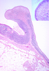
This is a low power view of a H&E stained slide showing normal parotid gland (salivary tissue) with serous acini in the upper half of the field adjacent to multi-loculated lesional tissue with cystic spaces lined by lymphoid tissue in which there is florid lymphoid hyperplasia with prominent germinal centres. The lumen of the cyst on the right contains haemorrhagic and keratinous debris including inflammatory cells and cholesterol clefts. The Inset (top right corner) is a medium power view of a H&E stained slide showing the epithelium lining the cyst wall which is mostly flattened squamous in type, showing the close relationship with the lymphoid stroma, as well as focal infiltration of lymphocytes into the epithelium.
Figure 2
Figure 2
This is a low power view of lesional tissue showing cystic spaces lined by squamous type epithelium with lymphoid tissue including a germinal centre on the right. These cysts show a mixture of squamous and ductal type epithelial lining with prominent infiltration by small lymphocytes. The Inset (top right corner) is low power view of a H&E stained slide showing ductal structures surrounded by blood vessels with abundant lymphoid tissue in the adjacent stroma. Scattered small islands of epithelium are identified in the lymphoid stroma. These represent branchial pouch-derived inclusions which proliferate to form cysts under the influence of growth factors produced by the hyperplastic lymphoid tissue. In line with the lymph node inclusion theory, some of these consist of pink staining oncocytic epithelium of the type as seen in Warthin’s tumours.F
Figure 3
Figure 3
The section shows neurovascular tissue at the top, adjacent to normal parotid salivary gland which in turn lies a cyst wall lined by bland epithelial cells with lymphoid tissue including a germinal centre in the wall. The Inset (top right corner) is medium power view of a cyst wall lined by bland squamous epithelium with abundant mixed lymphoid cells in the wall.
Figure 4
Figure 4
This shows vascular adipose tissue and nerve bundles underlying a cystic structure lined by bland squamous epithelium with lymphoid tissue in the wall. Also in the wall of the cyst is a ductal structure. The Inset (top right corner) shows a high power view of a structure lined by bland epithelial cells and fibrous tissue in the stroma.

References

    1. Hunczowski JN. Branchiogenetic or branchial fistulae. American Medicine. 1789;41:324.
    1. Langenbeck B. Exsterpation einer Dermoidcyste von der Scheide der grossen Halsgefasse. Verwundung der Vena jugularis communis. Stillung der blutung durch Compression Heilung. Archiv fur Klinische Chirurgie. 1859;1:25.
    1. Hildebrandt O. Uber angeborene epitheliale cysten und fisteln des halses. Archiv fur Klinische Chirurgie. 1895;49:167.
    1. Engzell U, Zujicek J. Aspiration biopsy of tumours of the neck I: Aspiration biopsy and cytological findings in 100 cases of congenital cysts. Acta Cytol. 1970;14:51. - PubMed
    1. Howie AJ, Proops DW. The definition of branchial cysts, sinuses and fistulae. Clin Otolaryngol. 1982;7:51–57. doi: 10.1111/j.1365-2273.1982.tb01561.x. - DOI - PubMed

LinkOut - more resources