Review
. 2012:87:219-47.
doi: 10.1016/B978-0-12-398312-1.00008-1.

Molecular modeling of inhibitors of human DNA methyltransferase with a crystal structure: discovery of a novel DNMT1 inhibitor

Affiliations
Review

Molecular modeling of inhibitors of human DNA methyltransferase with a crystal structure: discovery of a novel DNMT1 inhibitor

Jakyung Yoo et al. Adv Protein Chem Struct Biol. 2012.

Abstract

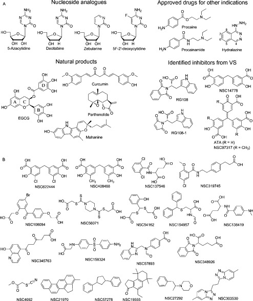
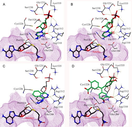
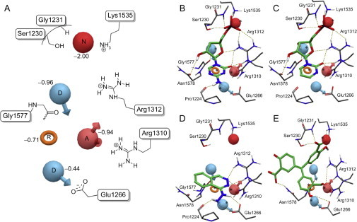
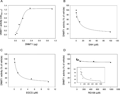
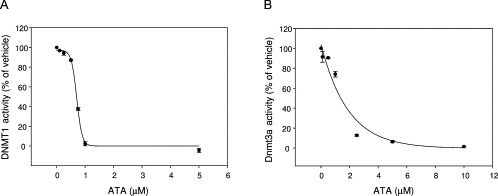
DNA methyltransferases (DNMTs) are promising epigenetic targets for the development of novel anticancer drugs and other diseases. Molecular modeling and experimental approaches are being used to identify and develop inhibitors of human DNMTs. Most of the computational efforts conducted so far with DNMT1 employ homology models of the enzyme. Recently, a crystallographic structure of the methyltransferase domain of human DNMT1 bound to unmethylated DNA was published. Following on our previous computational and experimental studies with DNMTs, we herein present molecular dynamics of the crystal structure of human DNMT1. Docking studies of established DNMT1 inhibitors with the crystal structure gave rise to a structure-based pharmacophore model that suggests key interactions of the inhibitors with the catalytic binding site. Results had a good agreement with the docking and pharmacophore models previously developed using a homology model of the catalytic domain of DNMT1. The docking protocol was able to distinguish active DNMT1 inhibitors from, for example, experimentally known inactive DNMT1 inhibitors. As part of our efforts to identify novel inhibitors of DNMT1, we conducted the experimental characterization of aurintricarboxylic acid (ATA) that in preliminary docking studies showed promising activity. ATA had a submicromolar inhibition (IC(50)=0.68 μM) against DNMT1. ATA was also evaluated for Dnmt3a inhibition showing an IC(50)=1.4 μM. This chapter illustrates the synergy from integrating molecular modeling and experimental methods to further advance the discovery of novel candidates for epigenetic therapies.


Figures

Fig. 1
Fig. 1
Mechanism of cytosine DNA methylation. Amino acid residue numbers are based on the crystal structure of hDNMT1. Equivalent residue numbers in parentheses correspond to the homology model.
Fig. 2
Fig. 2
Schematic representation of human DNMT1. NLS, nuclear localization signal; RFD, replication foci-targeting sequence; BAH, bromo-adjacent homology domain; TRD, target recognition domain. Interaction domains of HDAC1, HDAC2, and the DNMT3s are indicated. The methyltransferase domain comprising six most conserved motifs is enlarged.
Fig. 3
Fig. 3
(A) Methyltransferase domain of human DNMT1 (PDB: 3PTA) (pink). (B) Published structure of the M.HhaI–DNA complex (PDB: 1MHT) (yellow). (C) Previously developed homology model of DNMT1 (green). In (A)–(C), the catalytic loops are in black. Superposition of the crystal structure of human DNMT1 (1135–1600)–DNA complex with (D) the M.HhaI–DNA complex and (E) the homology model. The catalytic loop of the crystal structure is in red. AdoHcy and the flipped cytosine are shown in space-filling view. (F) Binding model of deoxycytidine (carbon atoms in black) with key amino acid residues of the crystal structure (carbon atoms in pink) and the homology model (carbon atoms in green). Hydrogen bonding interactions are depicted with dashes.
Fig. 4
Fig. 4
(A) Crystal structure of human DNMT1 (1135–1160)–DNA complex (gray) modified into an active state. The catalytic loop (residues 1224–1235) is highlighted with dark color. (B) Superposition of the modified crystal structure with the initial crystal structure of human DNMT1 (pink) and (C) homology model (green). The catalytic loops of the crystal structure and homology model are in red. AdoHcy and the flipped cytosine are shown in space-filling view. (D) Detail of the conformational change of the catalytic loop from “inactive” state (pink) into an active state (gray). (E) Binding interactions of modeled deoxycytidine (carbon atoms in black) with key amino acid residues in the modified crystal structure (carbon atoms in gray) and crystal structure (carbon atoms in pink). Hydrogen bonding interactions are depicted with dashes.
Fig. 5
Fig. 5
Compounds considered in this study that have been previously tested for inhibition of DNMT1: (A) active and (B) inactive/decoys.
Fig. 6
Fig. 6
Binding mode of representative inhibitors of DNMT1 (carbon atoms in green) into the catalytic site of the modified X-ray structure of DNMT1. The binding mode of deoxycytidine (carbon atoms in dark gray) is shown for reference: (A) 5-azacytidine, (B) zebularine, (C) hydralazine, (D) ATA.
Fig. 7
Fig. 7
(A) Structure-based pharmacophore model using binding modes of known inhibitors in the catalytic binding site of the modified crystal structure. Red sphere negative ionizable (N), pink sphere hydrogen bond acceptor (A), blue sphere hydrogen bond donors (D), and orange ring aromatic ring (R). Selected amino acid residues in the catalytic site are schematically depicted for reference. Comparison between the binding mode and pharmacophore hypothesis for representative DNMT inhibitors: (B) 5-azacytidine, (C) zebularine, (D) hydralazine, (E) ATA.
Fig. 8
Fig. 8
(A) Titration of recombinant DNMT1 enzymatic activity using a colorimetric assay. DNMT1 activity was measured using EpiQuik DNA methyltransferase activity/inhibition assay kit (Epigentek). (B–D) Dose–response plots for each compound against DNMT1. Data are presented in terms of percentage activity versus the results using vehicle only treated control, which was assigned a value of 100%. The IC50 concentrations of compounds were determined by enzyme assay under identical conditions (0.3 μg/1.6 pmol of DNMT1, incubated for 1 h).
Fig. 9
Fig. 9
(A) Dose–response plots for ATA inhibition against human DNMT1. Data are presented in terms of percent of vehicle only and the IC50 concentration of ATA was determined by enzyme assay with 0.3 μg of DNMT1 (1.6 pmol). (B) Dose–response plots for ATA inhibition against murine Dnmt3a. Data are presented in terms of percent of vehicle only control and the IC50 concentration of ATA was determined by enzyme assay with 4.0 μg of DNMT3A (33.3 pmol).

Similar articles

Cited by

References

    1. Arce C., Segura-Pacheco B., Perez-Cardenas E., Taja-Chayeb L., Candelaria M., Dueñnas-Gonzalez A. Hydralazine target: from blood vessels to the epigenome. J. Transl. Med. 2006;4:10. - PMC - PubMed
    1. Bouvet M., Debarnot C., Imbert I., Selisko B., Snijder E.J., Canard B. In vitro reconstitution of SARS-Coronavirus mRNA cap methylation. PLoS Pathog. 2010;6:e1000863. - PMC - PubMed
    1. Caulfield T., Medina-Franco J.L. Molecular dynamics simulations of human DNA methyltransferase 3B with selective inhibitor nanaomycin A. J. Struct. Biol. 2011;176:185–191. - PubMed
    1. Chen T.P., Hevi S., Gay F., Tsujimoto N., He T., Zhang B.L. Complete inactivation of DNMT1 leads to mitotic catastrophe in human cancer cells. Nat. Genet. 2007;39:391–396. - PubMed
    1. Cheng X.D., Blumenthal R.M. Mammalian DNA methyltransferases: a structural perspective. Structure. 2008;16:341–350. - PMC - PubMed

Publication types

MeSH terms

Substances