Review
. 2012 May 17;11(5):436-46.
doi: 10.1016/j.chom.2012.04.005.

Dendritic cells in antifungal immunity and vaccine design

Affiliations
Review

Dendritic cells in antifungal immunity and vaccine design

René M Roy et al. Cell Host Microbe. .

Abstract

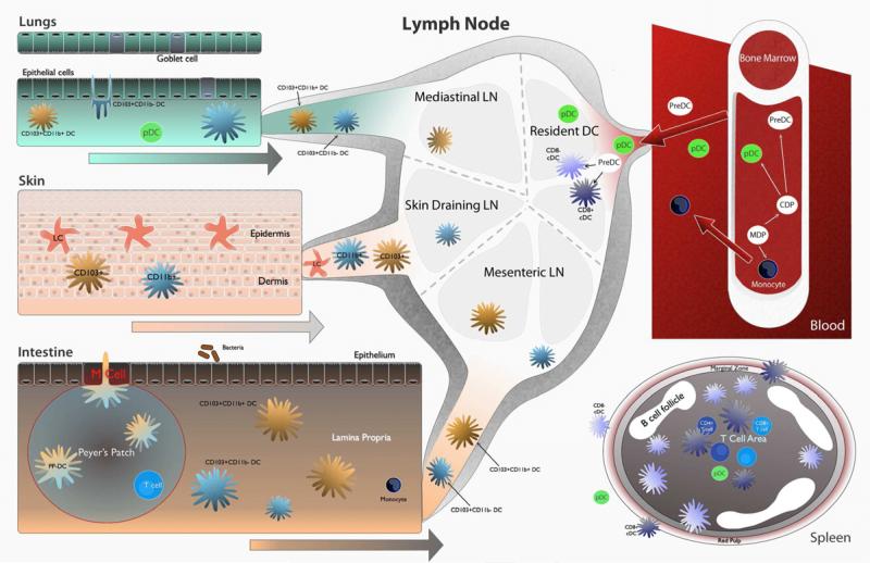
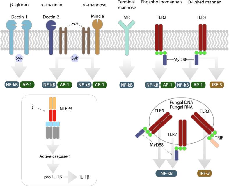
Life-threatening fungal infections have increased in recent years while treatment options remain limited. The development of vaccines against fungal pathogens represents a key advance sorely needed to combat the increasing fungal disease threat. Dendritic cells (DC) are uniquely able to shape antifungal immunity by initiating and modulating naive T cell responses. Targeting DC may allow for the generation of potent vaccines against fungal pathogens. In the context of antifungal vaccine design, we describe the characteristics of the varied DC subsets, how DC recognize fungi, their function in immunity against fungal pathogens, and how DC can be targeted in order to create new antifungal vaccines. Ongoing studies continue to highlight the critical role of DC in antifungal immunity and will help guide DC-based vaccine strategies.


Figures

Figure 1
Figure 1. Location of DC subsets important in anti-fungal immunity
DC subsets and monocytes arise from a common precursor in the bone marrow. pDC, monocytes, and PreDC exit the bone marrow and circulate via the blood. Resident CD8+ and CD8- DC exist in the spleen and the lymph node. PreDC seed the lungs, skin, and intestine and give rise to DC subsets in those locations. Migratory DC subsets migrate from peripheral locations to draining lymph nodes where they interact and prime T cells. In the lungs, CD103+ DC sample antigen from the airway lumen via paracellular processes. CD11b+ DC and CD103+ DC migrate to mediastinal lymph nodes. The epidermis of the skin contains a unique DC subset, Langerhans cells (LC) that are seeded in utero and self-renew. LC sample antigen and migrate via the dermis to skin draining lymph nodes. Dermal DC also migrate to skin draining lymph nodes. In the intestine, DC exist in the Peyer's Patch (PP) and the lamina propria (LP). PP-DC sample antigen using transcellular processes from the intestinal lumen following which they migrate to the T cell rich area of the PP where PP-DC prime T cells. LP-DC subsets migrate to the mesenteric lymph node following antigen sampling. During inflammation, monocytes enter inflamed tissue and differentiate into monocyte-derived DC (Mo-DC) which then carry antigen to draining lymph nodes.
Figure 2
Figure 2. Recognition of fungi via DC PRR
TLR and CLR. Fungal PAMP are recognized by several PRR: Dectin-1 signals via the tyrosine kinase Syk following recognition of β–glucan. Dectin-2 and Mincle, which also signal via Syk following recruitment of Fcγ, recognize alpha-mannan and alpha mannose respectively. Signaling downstream of the mannose receptor (MR) is undefined. TLR and CLR signaling results in downstream activation of the transcription factors NF-κB or AP-1. Some TLR signaling activates IRF3. While fungi activate the NLRP3 inflammasome, the mechanism is undefined. TLR recognize fungal carbohydrates, fungal DNA, and fungal RNA at the DC surface or in endosomal compartments.
Figure 3
Figure 3. DC-based strategies for developing anti-fungal vaccines
DC subsets may be targeted by varying the route of administration; aerosols target lung DC subsets, intradermal injection targets skin DC. DC can be loaded directly ex vivo before transfer into the host. Recombinant yeast contain ligands recognized by DC and allow for efficient DC uptake of antigen expressing organisms. β–glucan particles robustly activate DC via dectin-1. Virosomes also contain DC targeting ligands and viral PRR ligands that activate DC. Nanoparticles represent a complete engineered solution that incorporates PRR ligands, DC targeting ligands, and vaccine antigens. Following DC targeting, mature DC present antigen and activate naïve T cells.

Comment in

References

    1. Abdel-Motal UM, Berg L, Rosen A, Bengtsson M, Thorpe CJ, Kihlberg J, Dahmen J, Magnusson G, Karlsson KA, Jondal M. Immunization with glycosylated Kb-binding peptides generates carbohydrate-specific, unrestricted cytotoxic T cells. Eur J Immunol. 1996;26:544–551. - PubMed
    1. Allan RS, Waithman J, Bedoui S, Jones CM, Villadangos JA, Zhan Y, Lew AM, Shortman K, Heath WR, Carbone FR. Migratory dendritic cells transfer antigen to a lymph node-resident dendritic cell population for efficient CTL priming. Immunity. 2006;25:153–162. - PubMed
    1. Avci FY, Li X, Tsuji M, Kasper DL. A mechanism for glycoconjugate vaccine activation of the adaptive immune system and its implications for vaccine design. Nat Med. 2011;17:1602–1609. - PMC - PubMed
    1. Backer R, van Leeuwen F, Kraal G, den Haan JM. CD8- dendritic cells preferentially cross-present Saccharomyces cerevisiae antigens. Eur J Immunol. 2008;38:370–380. - PubMed
    1. Banchereau J, Steinman RM. Dendritic cells and the control of immunity. Nature. 1998;392:245–252. - PubMed

Publication types

Substances