. 2013 Jan;34(1):137-45.
doi: 10.1016/j.neurobiolaging.2012.04.007. Epub 2012 May 18.

Immunization targeting a minor plaque constituent clears β-amyloid and rescues behavioral deficits in an Alzheimer's disease mouse model

Affiliations

Immunization targeting a minor plaque constituent clears β-amyloid and rescues behavioral deficits in an Alzheimer's disease mouse model

Jose Morales-Corraliza et al. Neurobiol Aging. 2013 Jan.

Abstract

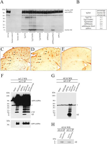
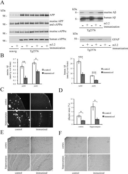
Although anti-human β-amyloid (Aβ) immunotherapy clears brain β-amyloid plaques in Alzheimer's disease (AD), targeting additional brain plaque constituents to promote clearance has not been attempted. Endogenous murine Aβ is a minor Aβ plaque component in amyloid precursor protein (APP) transgenic AD models, which we show is ∼3%-8% of the total accumulated Aβ in various human APP transgenic mice. Murine Aβ codeposits and colocalizes with human Aβ in amyloid plaques, and the two Aβ species coimmunoprecipitate together from brain extracts. In the human APP transgenic mouse model Tg2576, passive immunization for 8 weeks with a murine-Aβ-specific antibody reduced β-amyloid plaque pathology, robustly decreasing both murine and human Aβ levels. The immunized mice additionally showed improvements in two behavioral assays, odor habituation and nesting behavior. We conclude that passive anti-murine Aβ immunization clears Aβ plaque pathology--including the major human Aβ component--and decreases behavioral deficits, arguing that targeting minor endogenous brain plaque constituents can be beneficial, broadening the range of plaque-associated targets for AD therapeutics.


Figures

Figure 1
Figure 1. Co-deposition of murine and human Aβ in β-amyloid depositing mice
(A) Endogenous murine APP, sAPPα and Aβ levels in various APP and/or PS1 overexpressing tg lines and non-tg mice (see Materials and Methods for details) as detected by Western blot analysis of brain homogenates with antibody m3.2. Antibody m3.2 showed no reactivity with human control and AD brain tissue, in agreement with the specificity of this antibody for murine APP metabolites. (B) Percentage of murine Aβ of total Aβ (human + murine Aβ) accumulating in the brains of these tg lines as determined by ELISA. (C–E) Serial brain coronal sections were immunolabeled either with human-Aβ-specific antibody (C) or murine-Aβ-specific antibody (D) (with arrows marking co-labeled plaques) or with an addition of a 10-fold molar excess of murine Aβ to the murine-Aβ-specific antibody binding solution (E). (F–H) The murine-Aβ-specific antibody m3.2 was used to immunoprecipitate murine Aβ from brain homogenates of wild-type, APP ko, 16-month-old Tg2576 (depositing) and 4-month-old Tg2576 (predepositing) mice. m3.2-immunoprecipitation products were analyzed by Western blots probed with m3.2 for murine Aβ (F) or with 6E10 for human Aβ (G). The relative amount of human Aβ immunoprecipitated with 6E10 or co-immunoprecipitated with m3.2 is shown by 6E10 Western blot analysis (H). The asterisk (*) indicates non-specific reactivity.
Figure 2
Figure 2. Human and murine APP metabolite levels and Aβ plaque burden in the brains of Tg2576 mice following m3.2 immunotherapy
(A, left panels) Western blot analysis of total APP (both human and murine; detected by C1/6.1 from homogenate samples), both murine APP and sAPPα (detected by m3.2 from homogenate samples), murine sAPPα alone (detected by m3.2 from diethylamine (DEA)-extracted samples) and human sAPPα levels (detected by 6E10 from DEA-extracted samples) in brains of Tg2576 and non-tg mice injected with either m3.2 or control antibody. (A, right panels) Western blots of brain homogenates probed with m3.2 or 6E10 showing murine and human Aβ levels, respectively. Brain GFAP expression is shown by Western blot analysis in the bottom right panel. (B) ELISA measurement of formic acid-extracted murine Aβ40 and murine Aβ42 (left graph) and human Aβ40 and human Aβ42 (right graph) in Tg2576 mouse brains in m3.2-injected mice (n=7) compared to controls (n=6). (C) Thioflavin S staining of amyloid plaques in the cortex (upper panels) and hippocampus (lower panels) of Tg2576 mouse brains, comparing m3.2-injected (right panels) with control-antibody-injected mice (left panels). (D) Quantification of Thioflavin S positive plaque area in these mice (n=7, m3.2-injected mice and n=6, controls). Immunolabeling with anti-GFAP (E) and anti-ubiquitin (F) antibodies of representative brain coronal sections showing the cortex and hippocampus of m3.2-injected Tg2576 mice compared to control-antibody injected mice. *p<0.05; ** p<0.01; ***p<0.001.
Figure 3
Figure 3. Rescue of behavioral deficits in Tg2576 mice following m3.2 immunotherapy
(A) Odor habituation measurements of m3.2-injected Tg2576 mice (n=7) compared to control-injected Tg2576 mice (n=9). m3.2-injected (n=6) and control-injected (n=9) non-tg mice were also analyzed for comparison. (B) Nesting behavioural test analysis of the same groups of mice (see Supplemental Figure 1). *p<0.05; ** p<0.01; ***p<0.001.

References

    1. Bayer AJ, Bullock R, Jones RW, Wilkinson D, Paterson KR, Jenkins L, Millais SB, Donoghue S. Evaluation of the safety and immunogenicity of synthetic Aβ42 (AN1792) in patients with AD. Neurology. 2005;64(1):94–101. - PubMed
    1. Bodin K, Ellmerich S, Kahan MC, Tennent GA, Loesch A, Gilbertson JA, Hutchinson WL, Mangione PP, Gallimore JR, Millar DJ, Minogue S, Dhillon AP, Taylor GW, Bradwell AR, Petrie A, Gillmore JD, Bellotti V, Botto M, Hawkins PN, Pepys MB. Antibodies to human serum amyloid P component eliminate visceral amyloid deposits. Nature. 2010;468(7320):93–7. - PMC - PubMed
    1. Cataldo AM, Nixon RA. Enzymatically active lysosomal proteases are associated with amyloid deposits in Alzheimer brain. Proc Natl Acad Sci U S A. 1990;87(10):3861–5. - PMC - PubMed
    1. Chishti MA, Yang DS, Janus C, Phinney AL, Horne P, Pearson J, Strome R, Zuker N, Loukides J, French J, Turner S, Lozza G, Grilli M, Kunicki S, Morissette C, Paquette J, Gervais F, Bergeron C, Fraser PE, Carlson GA, George-Hyslop PS, Westaway D. Early-onset amyloid deposition and cognitive deficits in transgenic mice expressing a double mutant form of amyloid precursor protein 695. J Biol Chem. 2001;276(24):21562–70. - PubMed
    1. Choi-Miura NH, Ihara Y, Fukuchi K, Takeda M, Nakano Y, Tobe T, Tomita M. SP-40,40 is a constituent of Alzheimer's amyloid. Acta neuropathologica. 1992;83(3):260–4. - PubMed

Publication types

MeSH terms