. 2012 May 25;36(5):821-33.
doi: 10.1016/j.immuni.2012.03.021. Epub 2012 May 17.

Epithelial cell-specific Act1 adaptor mediates interleukin-25-dependent helminth expulsion through expansion of Lin(-)c-Kit(+) innate cell population

Affiliations

Epithelial cell-specific Act1 adaptor mediates interleukin-25-dependent helminth expulsion through expansion of Lin(-)c-Kit(+) innate cell population

Zizhen Kang et al. Immunity. .

Abstract

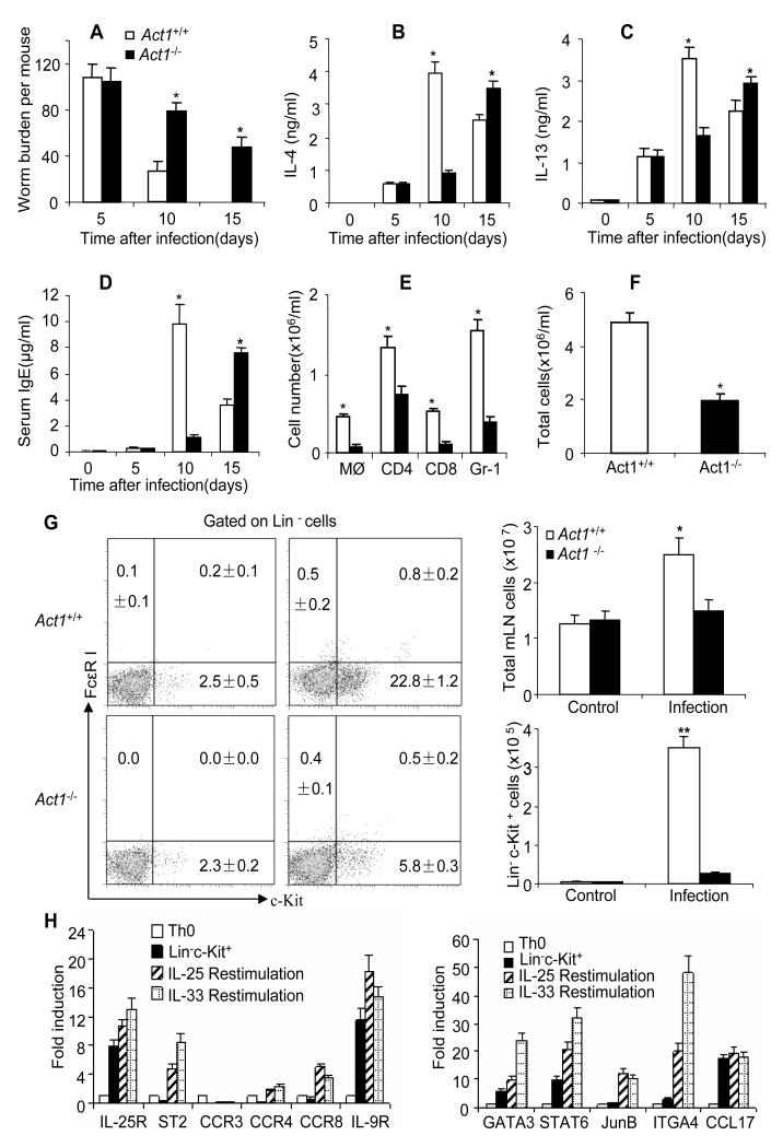
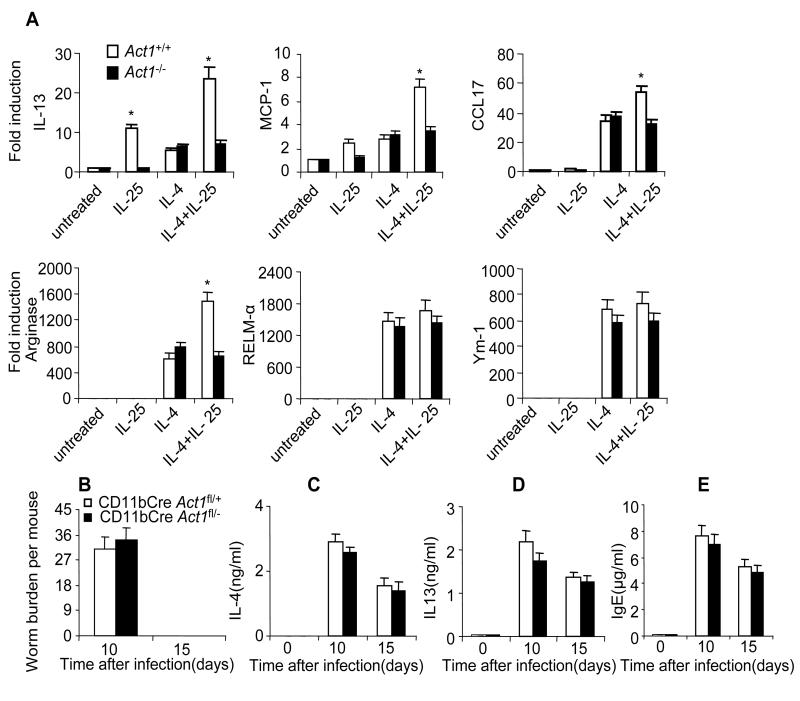
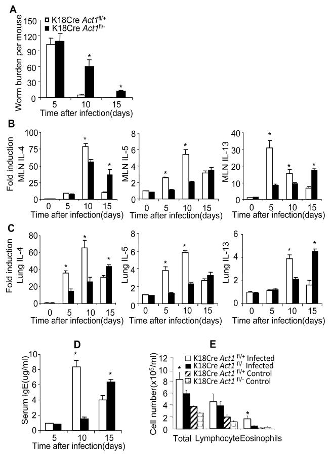
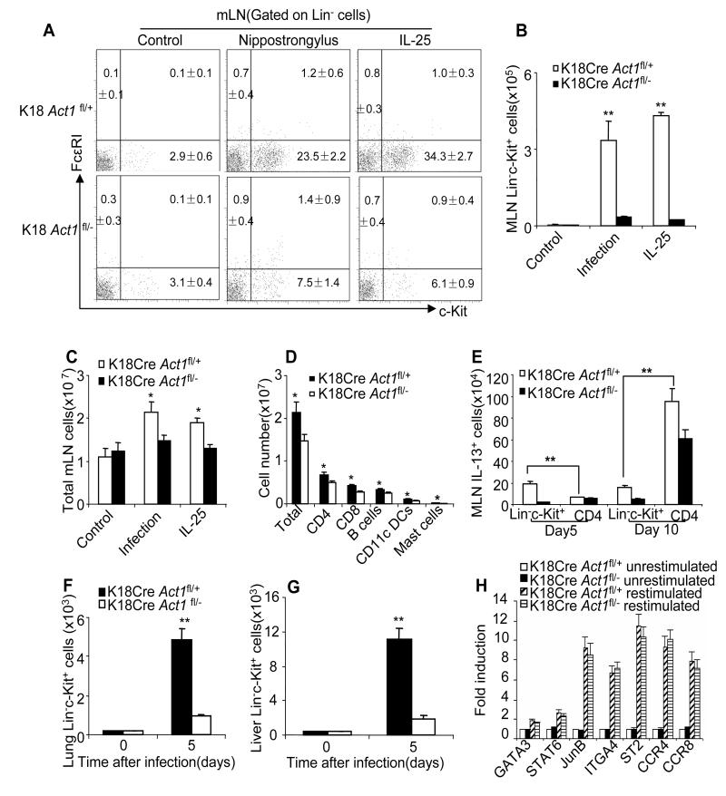
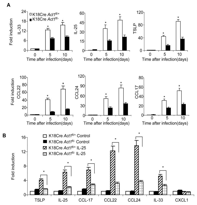
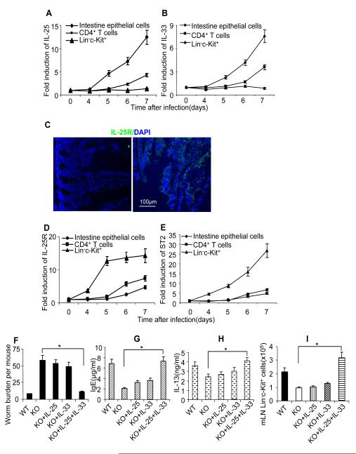
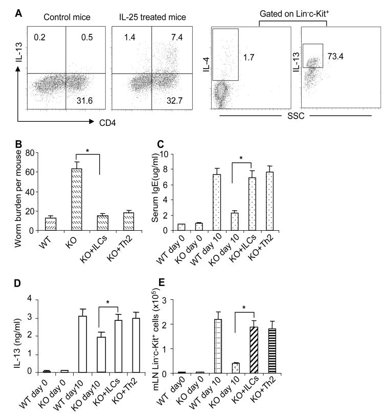
Interleukin-25 (IL-25 or IL-17E), a member of the structurally related IL-17 family, functions as an important mediator of T helper 2 cell-type (type 2) responses. We examined the cell type-specific role of IL-25-induced Act1-mediated signaling in protective immunity against helminth infection. Targeted Act1 deficiency in epithelial cells resulted in a marked delay in worm expulsion and abolished the expansion of the Lin(-)c-Kit(+) innate cell population in the mesenteric lymph node, lung, and liver. Th2 cell-inducing cytokine (IL-25 and IL-33) expression were reduced in the intestinal epithelial cells from the infected and IL-25-injected epithelial-specific Act1-deficient mice. Adoptive transfer of Lin(-)c-Kit(+) cells or combined injection of IL-25 and IL-33 restored the type 2 responses in these mice. Taken together, these results suggest that epithelial-specific Act1 mediates the expansion of the Lin(-)c-Kit(+) innate cell population through the positive-feedback loop of IL-25, initiating the type 2 immunity against helminth infection.


Figures

Figure 1
Figure 1. Act1 deficiency resulted in reduced type 2 responses and delayed worm expulsion
(A) Act1−/− and Act1+/+ mice were sacrificed 5, 10 and 15 days after infection, followed by analysis of worm burden in intestine of each mouse. (B-C) Mesenteric lymph nodes were taken from the infected mice at indicated time points. Cells were re-stimulated by anti-CD3 and anti-CD28 for overnight, followed by IL-4 and IL-13 ELISA in the collected supernatants. (D) Serum IgE levels were assayed by ELISA. (E-F) Different cell subsets in BAL of Act1-deficient and wild-type control mice were analyzed by flow cytometry 5 days after infection. Total cell numbers and absolute number of each subset were shown. (G) Act1-deficient and wild-type control mice were sacrificed 5 days after infection and mesenteric lymph nodes were collected. Plots shown are gated on live, lineage negative cells(CD3ε, CD4, CD8, TCRβ, TCRγδ, B220, CD19, NK1.1, Ter119, Gr-1, CD11b and CD11c), The frequency of Linc-kit+ cells(left panel) and absolute numbers were shown(right panel). All the data represent mean (n=4)±SEM; *p<0.05, **p<0.01. (H)Linc-kit+ cells were sorted by MACS and FACS, then sorted cells were restimulated by IL-25 or IL-33, Th0 helper T cells and unrestimulated Linc-kit+ cells were included as controls, specific gene expression was shown as mean(n=4)±SEM. The experiment was repeated three times. See also Figure S1;S2.
Figure 2
Figure 2. Macrophage Act1 is dispensable for worm expulsion
(A) Bone marrow macrophages from Act1−/− and Act1+/+ mice were untreated or treated with IL-25 (100ng/ml), IL-4 (10ng/ml) or IL-25+IL-4 for 24 hours, followed by analysis of gene expression by real-time PCR. (B) CD11bCre Act1fl/− mice and CD11bCre Act1fl/+ were sacrificed 10 and 15 days after infection, followed by analysis of worm burden in intestine of each mouse. (C-D) Mesenteric lymph nodes were taken from the infected mice at indicated time points. Cells were re-stimulated by anti-CD3 and anti-CD28 for overnight, followed by IL-4 and IL-13 ELISA in the collected supernatants. (E) Serum IgE levels were measured by ELISA. Data represent mean(n=4)±SEM; *p<0.05. The experiment was repeated three times.
Figure 3
Figure 3. Epithelial cell-specific Act1 is critical for type-2 dependent worm expulsion
(A) Epithelial-specific Act1-deficient and littermate control mice were sacrificed 10 and 15 days after infection, followed by analysis of worm burden in intestine of each mouse. (B-C) The expression of IL-4, IL-5 and IL-13 in the lung and mesenteric lymph nodes after infection were analyzed by real-time PCR. (D) Serum IgE concentrations were measured by ELISA. (E) BAL infiltration. Data represent mean(n=4)±SEM; *p<0.05. The experiment was repeated three times.
Figure 4
Figure 4. Act1-mediated signaling in epithelium is required for the IL-25-induced expansion of Linc-kit+ cells
(A) Frequencies of Linc-kit+ cells in the mesenteric lymph nodes of K18Cre Act1fl/− and K18Cre Act1fl/+ mice after 4 days of PBS or IL-25 treatment by i.p. or 5 days after worm infection. Plots shown are gated on lineage negative cells. (B-C) Total cell numbers (B) or Lin-c-kit+ cell numbers (C) in mesenteric lymph nodes from K18Cre Act1fl/− and K18Cre Act1fl/+ mice after 4 days of PBS or IL-25 treatment by i.p. or 5 days after worm infection. (D) Number of total MLN cells or of T cells(CD4+ or CD8+), B cells (CD19+), Dendritic cells(CD11c+) and mast cells from K18Cre Act1fl/− and K18Cre Act1fl/+ mice 5 days after worm infection. (E) Absolute number of MLN IL-13+ cells 5 or 10 days after infection. (F) Absolute number of Linc-kit+ cells in the lung. (G) Absolute number of Lin-c-kit+ cells in the liver. (H) Gene expression of MLN Linc-kit+ cells from K18Cre Act1fl/− and K18Cre Act1fl/+ mice 5 days after worm infection. Isolated cells were restimulated with IL-25, and cells without restimulation were included as controls. Data represent mean(n=4)±SEM; *p<0.05, **p<0.01. The experiment was repeated two times. See also Figure S3.
Figure 5
Figure 5. IL-25 induced type 2 cytokines and chemokines in epithelial cells
(A) Infected K18Cre Act1fl/− and K18Cre Act1fl/+ mice were sacrificed 10 days after infection. Gene expressions in intestinal epithelial cells after infection were analyzed by real-time PCR. (B) Epithelial-specific Act1-deficient and littermate control mice were subjected to intra-peritoneal injection of IL-25 (2μg/mouse, every 24 hours for two days), followed by analysis of gene expression in intestinal epithelial cells by real-time PCR. Data represent mean(n=4)±SEM; *p<0.05. The experiment was repeated two times.
Figure 6
Figure 6. Combined injection of IL-25 and IL-33 restored type 2 immunity in epithelial-specific Act1-deficient mice
Wild type mice were infected with N. brasiliensis, mice were sacrificed at different time points as indicated and CD4+ T cells, Linc-kit+ cells and intestinal epithelial cells were isolated and gene expression were analyzed by real-time PCR. (A) IL-25 expression. (B) IL-33 expression. (C) cross sections of small intestine from naïve C57BL/6 mice were stained with anti-IL-25R antibodies(Right), isotype antibody staining was included as control(left). Green fluorescence suggests IL-25R positive. Blue shows DAPI staining. (D) IL-25R expression (samples from A-B). (E) ST2 expression (samples from A-B). K18Cre Act1fl/−(KO) and K18Cre Act1fl/+ (WT) mice were infected(F-I), three groups of KO mice received IL-25, IL-33 and IL-25+IL-33 injection (i.p, 500ng/mouse for 5 days starting from the first day of infection), respectively. All the mice were sacrificed 10 days after infection. (F) worm burden in small intestine was analyzed. (G) serum IgE levels were measured by ELISA. H. MLN Cells were re-stimulated by anti-CD3 and anti-CD28 for overnight, followed by IL-13 ELISA in the collected supernatants. I. Absolute numbers of Linc-kit+ cells in MLN are shown. Data represent mean(n=4)±SEM; *p<0.05. The experiment was repeated two times.
Figure 7
Figure 7. IL-13-producing cells rescued type 2 protective immunity in epithelial-specific Act1-deficient mice
(A) Th2 cells were induced by i.p injection of IL-25 consecutively for 7 days at a dose of 1000 ng/mouse each day. Linc-Kit+ cells were induced by i.p injection of IL-25 consecutively for 5 days at a dose of 500 ng/mouse each day. The frequencies of IL-13-producing CD4+ T cells and Linc-Kit+ cells are shown. K18Cre Act1fl/−(KO) and K18Cre Act1fl/+ (WT) mice were infected(B-E), one group of KO mice received 1×10⁶ Linc-kit+ cells/mouse and another group of KO mice received 107 CD4+ T cells/mouse at the same day of infection. All the mice were sacrificed 10 days after infection. (B)worm burden in small intestine were analyzed. (C) Serum IgE levels were measured by ELISA. (D) MLN Cells were re-stimulated by anti-CD3 and anti-CD28 for overnight, followed by IL-13 ELISA in the collected supernatants. (E) Absolute numbers of Linc-kit+ cells in MLN are shown. Data represent mean(n=4)±SEM; *p<0.05. The experiment was repeated two times. See also Figure S4.

Comment in

References

    1. Angkasekwinai P, Park H, Wang YH, Wang YH, Chang SH, Corry DB, Liu YJ, Zhu Z, Dong C. Interleukin 25 promotes the initiation of proallergic type 2 responses. J Exp Med. 2007;204:1509–1517. - PMC - PubMed
    1. Anthony RM, Rutitzky LI, Urban JF, Jr., Stadecker MJ, Gause WC. Protective immune mechanisms in helminth infection. Nat Rev Immunol. 2007;7:975–987. - PMC - PubMed
    1. Artis D. Epithelial-cell recognition of commensal bacteria and maintenance of immune homeostasis in the gut. Nat Rev Immunol. 2008;8:411–420. - PubMed
    1. Boillee S, Yamanaka K, Lobsiger CS, Copeland NG, Jenkins NA, Kassiotis G, Kollias G, Cleveland DW. Onset and progression in inherited ALS determined by motor neurons and microglia. Science. 2006;312:1389–1392. - PubMed
    1. Chang SH, Dong C. Signaling of interleukin-17 family cytokines in immunity and inflammation. Cell Signal. 2011;23:1069–1075. - PMC - PubMed

Publication types

MeSH terms