

Aquaporin 4 and neuromyelitis optica
- PMID: 22608667
- PMCID: PMC3678971
- DOI: 10.1016/S1474-4422(12)70133-3
Aquaporin 4 and neuromyelitis optica
Abstract
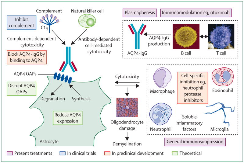
Neuromyelitis optica is an inflammatory demyelinating disorder of the CNS. The discovery of circulating IgG1 antibodies against the astrocyte water channel protein aquaporin 4 (AQP4) and the evidence that AQP4-IgG is involved in the development of neuromyelitis optica revolutionised our understanding of the disease. However, important unanswered questions remain--for example, we do not know the cause of AQP4-IgG-negative disease, how astrocyte damage causes demyelination, the role of T cells, why peripheral AQP4-expressing organs are undamaged, and how circulating AQP4-IgG enters neuromyelitis optica lesions. New drug candidates have emerged, such as aquaporumab (non-pathogenic antibody blocker of AQP4-IgG binding), sivelestat (neutrophil elastase inhibitor), and eculizumab (complement inhibitor). Despite rapid progress, randomised clinical trials to test new drugs will be challenging because of the small number of individuals with the disorder.
Copyright © 2012 Elsevier Ltd. All rights reserved.
Conflict of interest statement
The University of California San Francisco and the University of Colorado have filed a joint US provisional patent application for aquaporumab antibody treatment for neuromyelitis optica, of which ASV is a named co-inventor. The intellectual property rights are owned by the University of California and the University of Colorado. MCP declares that he has no conflicts of interest.
Figures
References
-
- Jarius S, Wildemann B. ‘Noteomielite’ accompanied by acute amaurosis (1844): an early case of neuromyelitis optica. J Neurol Sci. 2012;313:182–84. - PubMed
-
- Jarius S, Wildemann B. The case of the Marquis de Causan (1804): an early account of visual loss associated with spinal cord inflammation. J Neurol Sci. 2012;313:182–84. - PubMed
-
- Jarius S, Wildemann B. An early case of neuromyelitis optica: on a forgotten report by Jacob Lockhart Clarke, FRS. Mult Scler. 2011;17:1384–86. - PubMed
-
- Lennon VA, Wingerchuk DM, Kryzer TJ, et al. A serum autoantibody marker of neuromyelitis optica: distinction from multiple sclerosis. Lancet. 2004;364:2106–12. - PubMed
Publication types
MeSH terms
Substances
Grants and funding
LinkOut - more resources
Full Text Sources
Other Literature Sources
