. 2012 Aug;143(2):408-17.e2.
doi: 10.1053/j.gastro.2012.05.007. Epub 2012 May 15.

Neuronal serotonin regulates growth of the intestinal mucosa in mice

Affiliations

Neuronal serotonin regulates growth of the intestinal mucosa in mice

Erica R Gross et al. Gastroenterology. 2012 Aug.

Erratum in

  • Gastroenterology. 2013 Jan;144(1):249. Li, Zhishan [added]

Abstract

Background & aims: The enteric abundance of serotonin (5-HT), its ability to promote proliferation of neural precursors, and reports that 5-HT antagonists affect crypt epithelial proliferation led us to investigate whether 5-HT affects growth and maintenance of the intestinal mucosa in mice.

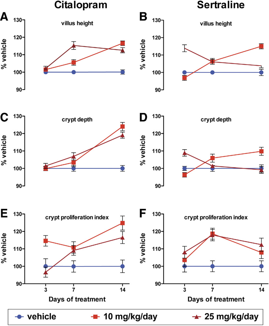
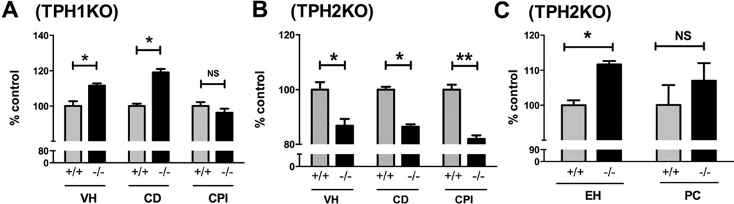
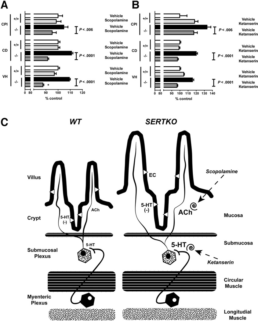
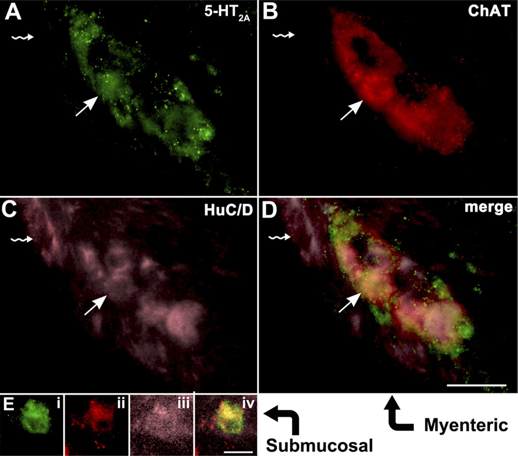
Methods: cMice that lack the serotonin re-uptake transporter (SERTKO mice) and wild-type mice were given injections of selective serotonin re-uptake inhibitors (gain-of-function models). We also analyzed mice that lack tryptophan hydroxylase-1 (TPH1KO mice, which lack mucosal but not neuronal 5-HT) and mice deficient in tryptophan hydroxylase-2 (TPH2KO mice, which lack neuronal but not mucosal 5-HT) (loss-of-function models). Wild-type and SERTKO mice were given ketanserin (an antagonist of the 5-HT receptor, 5-HT(2A)) or scopolamine (an antagonist of the muscarinic receptor). 5-HT(2A) receptors and choline acetyltransferase were localized by immunocytochemical analysis.

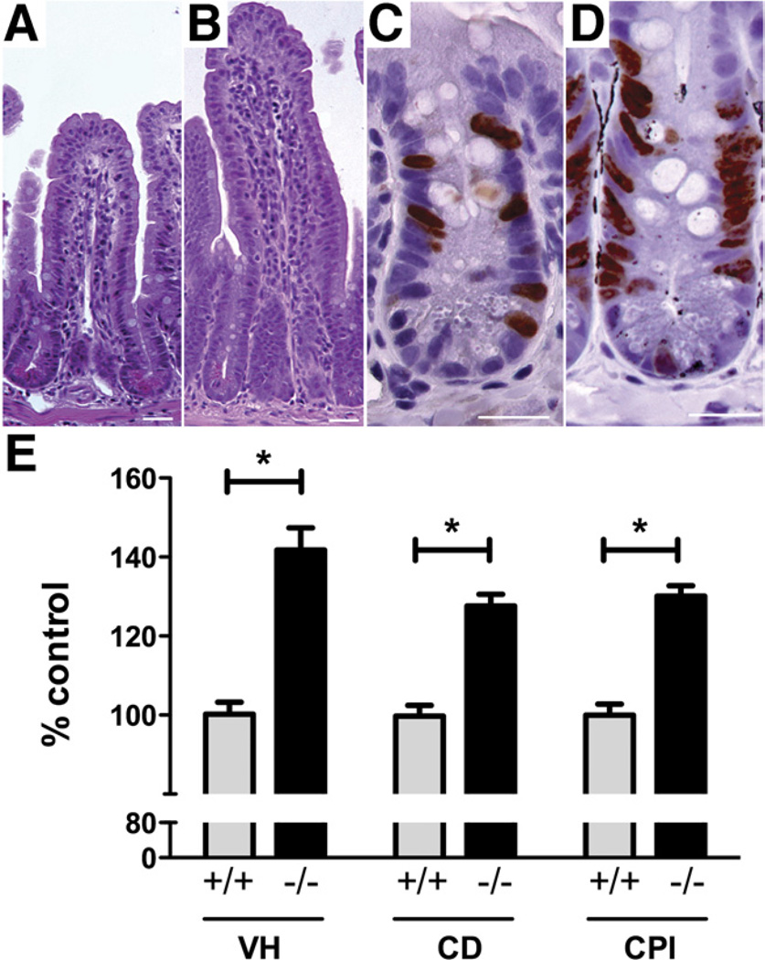
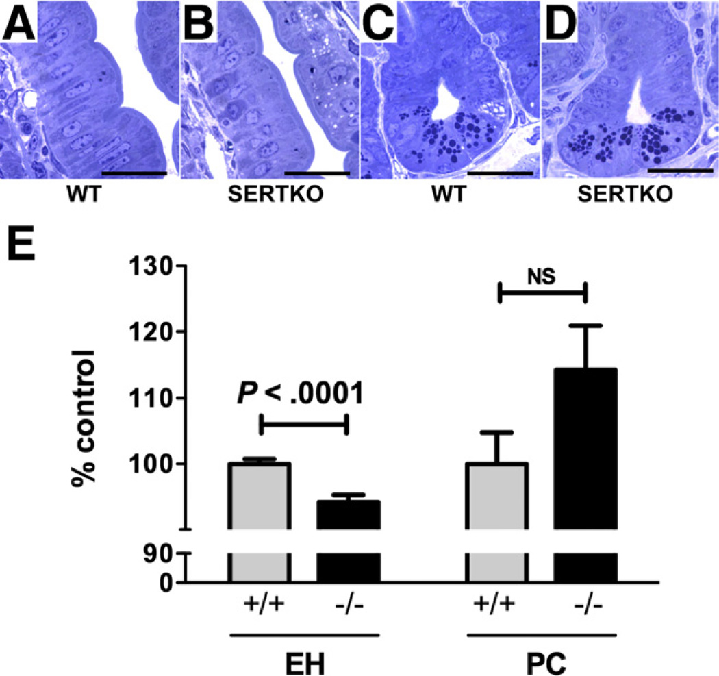
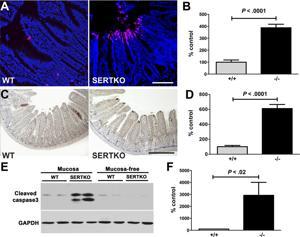
Results: Growth of the mucosa and proliferation of mucosal cells were significantly greater in SERTKO mice and in mice given selective serotonin re-uptake inhibitors than in wild-type mice, but were diminished in TPH2KO (but not in TPH1KO) mice. Ketanserin and scopolamine each prevented the ability of SERT knockout or inhibition to increase mucosal growth and proliferation. Cholinergic submucosal neurons reacted with antibodies against 5-HT(2A).

Conclusions: 5-HT promotes growth and turnover of the intestinal mucosal epithelium. Surprisingly, these processes appear to be mediated by neuronal, rather than mucosal, 5-HT. The 5-HT(2A) receptor activates cholinergic neurons, which provide a muscarinic innervation to epithelial effectors.


Conflict of interest statement

Conflicts of interest

These authors disclose the following: Kara Margolis and Michael Gershon received research support from Lexicon Genetics, and Michael Gershon is also a consultant for The Dannon Company, Inc. The remaining authors disclose no conflicts.

Figures

Figure 1
Figure 1
VH, CD, and CPI are significantly greater in SERTKO than in WT mice. (A) Mucosal architecture in a section of WT mouse ileum stained with H&E. (B) Mucosal architecture of a SERTKO mouse. (C) BrdU incorporation (brown reaction product) into crypt cells from a WT mouse. (D) BrdU incorporation in a SERTKO mouse. (A–D) Scale bars: 30 µm. (E) VH, CD, and CPI are expressed as the percentage of control (WT). *P < .0001.
Figure 2
Figure 2
Enterocytes are smaller in SERTKO than in WT mice. (A) Toluidine blue–stained semithin sections (0.7 µm). (A) Midvillus, WT. (B) Midvillus, SERTKO. (C) Crypt, WT. (D) Crypt, SERTKO. (A–D) Scale bars: 20µm. (E) EH and Paneth cell (PC) number are quantified and expressed as the percentage of control (WT).
Figure 3
Figure 3
Apoptosis of enterocytes at villus tips is greater in SERTKO than in WT mice. (A) Apoptotic cells (red) shown with TUNEL in the epithelium of the intestines of WT and SERTKO mice. DNA counterstained with bisbenzimide. Scale bar: 100 µm. (B) TUNEL staining cells quantified and expressed as the percentage of control (WT). (C) Cleaved caspase-3 immunostaining (brown). Scale bar: 200 µm. (D) Numbers of cells displaying cleaved caspase-3 immunostained cells quantified and expressed as the percentage of control (WT). (E) Western blot showing cleaved caspase-3 immunoreactivity in preparations of mucosa and the mucosa-free bowel wall in WT and SERTKO mice. GAPDH, glyceraldehyde-3-phosphate dehydrogenase. (F) Cleaved caspase-3 immunoreactivity was quantified in the mucosa of WT and SERTKO mice and expressed as the percentage of control (WT).
Figure 4
Figure 4
VH, CD, and CPI in WT mice that received vehicle (saline or 50% ETOH), citalopram (10 or 25 mg/kg/day), or sertraline (10 or 25 mg/kg/day) for 3, 7, or 14 days.
Figure 5
Figure 5
(A and B) VH, CD, and CPI have been quantified and expressed as the percentage of control (WT). (A) VH and CD, but not CPI, are significantly greater in TPH1KO than in WT mice. *P < . 0001, NS = 0. 2431. (B) VH, CD, and CPI are significantly lower in TPH2KO than in WT mice. (B) *P < . 0001, **P < . 01. (C) The height of enterocytes but not the numbers of Paneth cells in TPH2KO was greater than in WT mice. *P < . 05.
Figure 6
Figure 6
(A) VH, CD, and CPI are all significantly shorter in SERTKO mice treated with scopolamine than with vehicle. VH, CD, and CPI were quantified and expressed as the percentage of control (WT + vehicle). (B) VH, CD, and the CPI are all significantly shorter in SERTKO mice treated with ketanserin than with vehicle. (C) A hypothetical explanation of the mucosal changes seen in SERTKO mice. The deletion of SERT amplifies the effects of 5-HT that neurons secrete. Serotonergic stimulation of 5-HT2A–receptor– expressing cholinergic neurons in submucosal ganglia causes release of ACh in the mucosa, which stimulates epithelial growth. Blocking muscarinic receptors with scopolamine or 5-HT2A receptors with ketanserin thus prevent SERTKO-associated mucosal growth.
Figure 7
Figure 7
Cholinergic neurons in both the myenteric and submucosal plexuses express 5-HT2A immunoreactivity. (A–D) Myenteric ganglion. (A) 5-HT2A immunoreactivity. (B) ChAT immunoreactivity. (C) HuC/D immunoreactivity. (D) Merged image. The arrows depict a neuron that is triply labeled by all 3 markers. The arrows with a rippled stem depict a neuron that is not cholinergic and expresses neither 5-HT2A nor ChAT. (Ei–Eiv) Neuron of the submucosal plexus (neurons tend to occur singly in sections through the murine submucosal plexus). Ei, 5-HT2A immunoreactivity; Eii, ChAT immunoreactivity; Eiii, HuC/D immunoreactivity; and Eiv, merged image. Scale bars: 20 µm.

References

    1. Gershon MD, Tack J. The serotonin signaling system: from basic understanding to drug development for functional GI disorders. Gastroenterology. 2007;132:397–414. - PubMed
    1. Furness JB. The enteric nervous system. Malden, MA: Blackwell Publishing; 2006.
    1. Erspamer V. Pharmacology of indolealklyamines. Pharmacol Rev. 1954;6:425–487. - PubMed
    1. Vialli M. Histology of the enterochromaffin cell system. In: Erspamer V, editor. Handbook of experimental pharmacology: 5-hydroxytryptamine and related indolealkylamines. Volume 19. New York: Springer-Verlag; 1966. pp. 1–65.
    1. Gershon MD, Drakontides AB, Ross LL. Serotonin: synthesis and release from the myenteric plexus of the mouse intestine. Science. 1965;149:197–199. - PubMed

Publication types

MeSH terms