. 2012 Sep;1818(9):2282-9.
doi: 10.1016/j.bbamem.2012.05.013. Epub 2012 May 17.

Targeting the lateral interactions of transmembrane domain 5 of Epstein-Barr virus latent membrane protein 1

Affiliations

Targeting the lateral interactions of transmembrane domain 5 of Epstein-Barr virus latent membrane protein 1

Xiaohui Wang et al. Biochim Biophys Acta. 2012 Sep.

Abstract

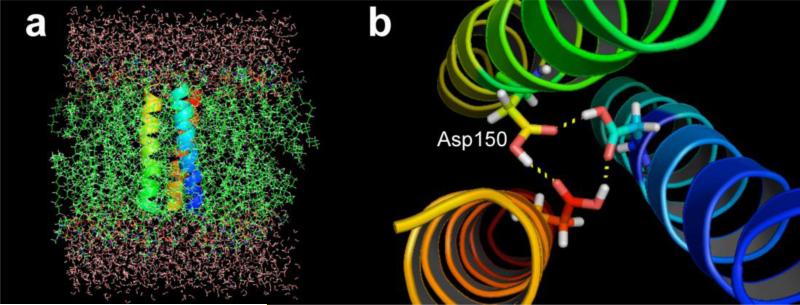
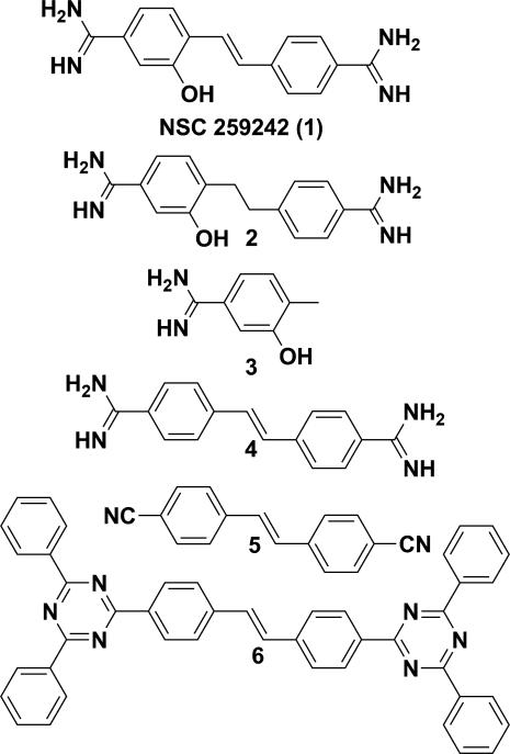
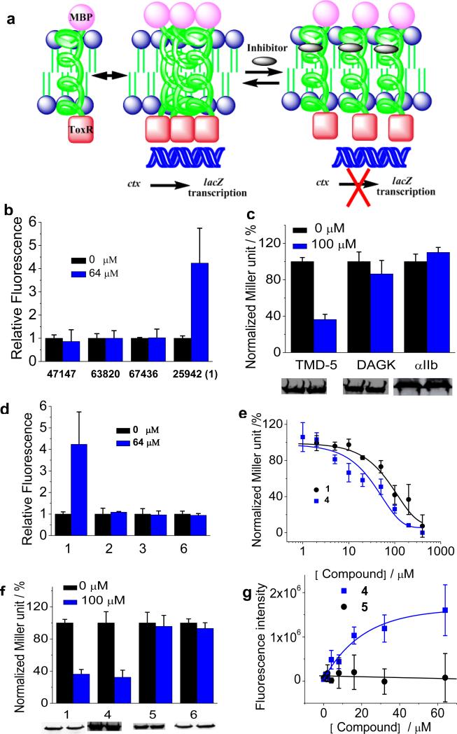
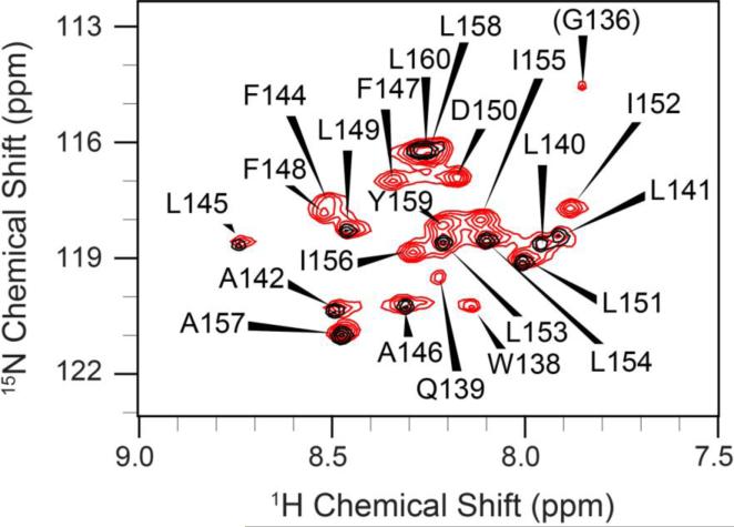
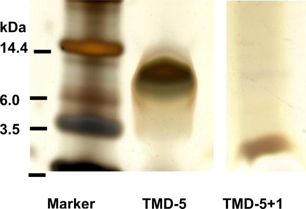
The lateral transmembrane protein-protein interaction has been regarded as "undruggable" despite its importance in many biological processes. The homo-trimerization of transmembrane domain 5 (TMD-5) of latent membrane protein 1 (LMP-1) is critical for the constitutive oncogenic activation of the Epstein-Barr virus (EBV). Herein, we report a small molecule agent, NSC 259242 (compound 1), to be a TMD-5 self-association disruptor. Both the positively charged acetimidamide functional groups and the stilbene backbone of compound 1 are essential for its inhibitory activity. Furthermore, cell-based assays revealed that compound 1 inhibits full-length LMP-1 signaling in EBV infected B cells. These studies demonstrated a new strategy for identifying small molecule disruptors for investigating transmembrane protein-protein interactions.


Figures

Figure 1
Figure 1
(a), Energy-optimized atomistic model [9] of homo-trimeric complex of LMP-1 TMD-5 in explicit membrane bilayers. (b), The TMD-5 association is driven by the H-bonds mediated by Asp 150.
Figure 2
Figure 2
Structure of NSC 259242 (compound 1) and its analogues.
Figure 3
Figure 3
(a), Schematic representation of the ToxR assay used for screening LMP-1 TMD-5 disruptors. (b), Coumarin fluorescence dequeching assay. Compounds were added into 100 nM of coumarin-labeled TMD-5 solution (50 mM HEPES, 150 μM C14 betaine, pH = 7.4) and equilibrated overnight. Samples were excited at 360 nm and emission was read at 430 nm using a Beckman-Coulter DTX 880 Multimode Detector plate reader. The background correction and the fluorescence of inhibitors were subtracted from the observed coumarin fluorescence signal. The fluorescence intensity of control sample (no compound) was set as 1.0. (c), ToxR measurement of the inhibitory effect of compound 1 on the oligomerization of LMP-1 TMD-5 and TMDs of diacylglycerol kinase (DAGK) and integrin αIIb. TMD oligomerization activity in the absence of compound was normalized as 100%. Western blot showed the chimeric MBP-TMD-ToxR protein expression level and gel loading normalized by OD600 nm of cultures. (d), Fluorescence dequenching assay of compound 1 and its analogues. Experiments were carried out as described in (b). (e), Dose-dependent titration of 1 and 4 on TMD-5 oligomerization using the ToxR assay. (f), ToxR measurement of oligomerization of TMD-5 in the presence of compound 1 or its analogues. Western blot showed the chimeric MBP-TMD-5-ToxR protein expression level and gel loading normalized by OD600 nm of cultures. (g), the dose dependent fluorescence enhancement curves of coumarin-labeled TMD-5 induced by compound 4 and compound 5. Experiments were carried out as described in (b).
Figure 4
Figure 4
2D 1H, 15N-HSQC spectra(excluding most the Gly region) of the TMD-5 peptides solubilized in bicelles. Red and black contours correspond to the spectrum of uniformly 13C, 15N enriched TMD-5 and selectively 15N-GLA labeled TMD-5, respectively. The spectrum shows nice chemical shift dispersion with well-defined peaks and line shapes, indicative of a folded, homogenous protein tertiary structure. Residues are numbered according their position in the native LMP-1 sequence.
Figure 5
Figure 5
The chemical shift index (CSI) values of (a) Cα and (b) CO of residues in LMP-1 TMD-5 confirmed that the TMD-5 peptide adopts a native α-helical conformation under the condition used in the NMR experiments
Figure 6
Figure 6
12% Bis-Tris SDS-PAGE analysis of TMD-5 after NMR titration. The ratio of compound 1 to TMD-5 in TMD-5 + 1 sample shown here is 4:1. Compound 1 disrupts the TMD-5 trimer (MW 10.2 kDa ) and causes TMD-5 monomer (3.4 kDa) formation. It should be noted that the loading amount of TMD-5 in lane 2 was greater than that of lane 3.
Figure 7
Figure 7
(a), Dose-dependent inhibitory effects of the full-length LMP-1-mediated NF-κB activity in EBV-infected B cells by TMD-5 inhibitor 1 and its analogues. (b), Effects of compound 1 and analogues on the NF-κB downstream signaling of NO production. (c), Cell proliferation assay. No apparent growth inhibition was observed at a concentration 100 μM of compounds 1 and its analogues. (d), Kinases profile of compound 1, (50 μM) measured by KinaseSeeker™ assay. Staurosporine (10 μM) was served as the positive control for kinase inhibitor.

Similar articles

Cited by

References

    1. Sackett DL, Sept D. Protein-protein interactions: making drug design second nature. Nat. Chem. 2009;1:596–597. - PubMed
    1. Arkin MR, Wells JA. Small-molecule inhibitors of protein-protein interactions: progressing towards the dream. Nat. Rev. Drug Discov. 2004;3:301–317. - PubMed
    1. Wilson AJ. Inhibition of protein-protein interactions using designed molecules. Chem. Soc. Rev. 2009;38:3289–3300. - PubMed
    1. Yin H. Exogenous agents that target transmembrane domains of proteins. Angew. Chem. Int. Ed. Engl. 2008;47:2744–2752. - PubMed
    1. Zhao TX, Martinko AJ, Le VH, Zhao J, Yin H. Development of agents that modulate protein-protein interactions in membranes. Curr. Pharm. Des. 2010;16:1055–1062. - PubMed

Publication types

MeSH terms