Review
. 2012 Oct 11;527(2):90-9.
doi: 10.1016/j.neulet.2012.04.078. Epub 2012 May 17.

Gene therapy for muscular dystrophy: lessons learned and path forward

Affiliations
Review

Gene therapy for muscular dystrophy: lessons learned and path forward

Jerry R Mendell et al. Neurosci Lett. .

Abstract

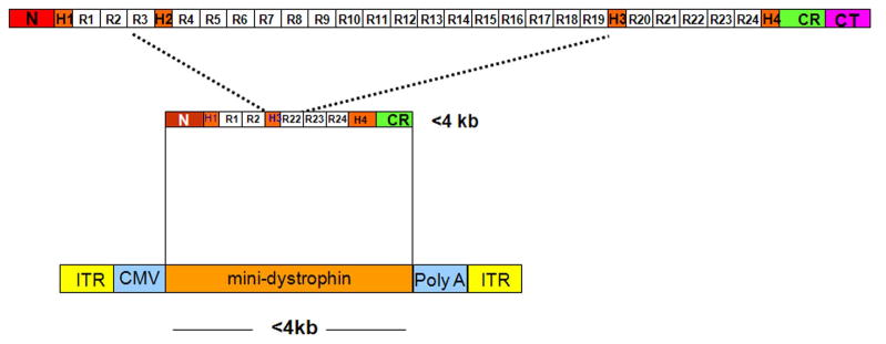
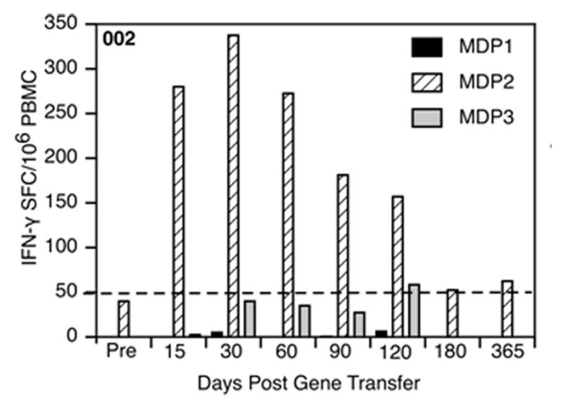
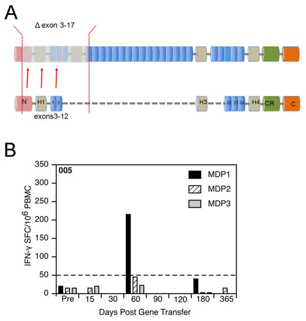
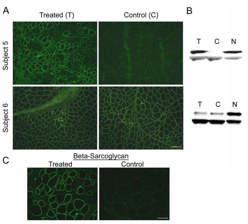
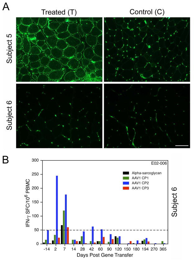
Our Translational Gene Therapy Center has used small molecules for exon skipping and mutation suppression and gene transfer to replace or provide surrogate genes as tools for molecular-based approaches for the treatment of muscular dystrophies. Exon skipping is targeted at the pre-mRNA level allowing one or more exons to be omitted to restore the reading frame. In Duchenne Muscular Dystrophy (DMD), clinical trials have been performed with two different oligomers, a 2'O-methyl-ribo-oligonucleoside-phosphorothioate (2'OMe) and a phosphorodiamidate morpholino (PMO). Both have demonstrated early evidence of efficacy. A second molecular approach involves suppression of stop codons to promote readthrough of the DMD gene. We have been able to establish proof of principle for mutation suppression using the aminoglycoside, gentamicin. A safer, orally administered, alternative agent referred to as Ataluren (PTC124) has been used in clinical trials and is currently under consideration for approval by the FDA. Using a gene therapy approach, we have completed two trials and have initiated a third. For DMD, we used a mini-dystrophin transferred in adeno-associated virus (AAV). In this trial an immune response was seen directed against transgene product, a quite unexpected outcome that will help guide further studies. For limb girdle muscular dystrophy 2D (alpha-sarcoglycan deficiency), the transgene was again transferred using AAV but in this study, a muscle specific creatine kinase promoter controlled gene expression that persisted for six months. A third gene therapy trial has been initiated with transfer of the follistatin gene in AAV directly to the quadriceps muscle. Two diseases with selective quadriceps muscle weakness are undergoing gene transfer including sporadic inclusion body myositis (sIBM) and Becker muscular dystrophy (BMD). Increasing the size and strength of the muscle is the goal of this study. Most importantly, no adverse events have been encountered in any of these clinical trials.


Figures

Figure 1
Figure 1. Full dystrophin protein
Amino-terminal (N), cysteine-rich (CR) domain, C-terminal (CT), and rod domain seen with 24 spectrin repeats (R1–R24) and four hinges (H1, H2, H3, H4). The mini-dystrophin represents a truncated peptide with N-terminal, CR domain and rod domain with reduced number of spectrin repeats (R1, R2, R22, R23, R24) and 3 hinges (H1, H3, H4). The mini-dystrophin cDNA with its Poly A tail is under control of the cytomegalovirus (CMV) promoter flanked by inverted terminal repeats (ITRs).
Figure 2
Figure 2. T cell responses clinical DMD gene therapy trial
Peripheral blood mononuclear cells (PBMC) were collected on the indicated days and cultured with three pools of synthetic peptides (MDP1, MDP2, MDP3) spanning the mini-dystrophin expressed by the transgene. Interferon-γ production was assessed 36 hours later with use of an interferon-γ enzyme-linked immunosorbent spot (ELISPOT) assay. Data are presented as interferon-γ spot forming cells (SFC) per 1 million PBMCs. The dashed line represents the threshold for a positive assay response (50 SFCs per 1 million PBMCs).
Figure 3
Figure 3. Patient’s deleted dystrophin peptide and transgene product
A) Depiction of dystrophin peptide resulting from deletion of exons 3 to 17 of DMD gene. Below is the truncated peptide translated from mini-dystrophin. The peptide expresses in the region of the patient’s deletion. The consequences are shown in B) where an immune response is demonstrated by ELISPOT assay to mini-dystrophin peptide 1 (MDP-1) composed of amino acids (aa) 1–469. There are no T cell responses generated by culturing with MDP2 or MDP3.
Figure 4
Figure 4. ELISPOT assay pre- and post-gene therapy
A) T cell response of PBMCs is stimulated by an amino acid fragment translated by exon 57. There is a robust T cell response seen post-treatment and minimal but suggestive immune response even pre-treatment. B) Revertant fiber cluster (3 positive fibers) is seen using C-terminal antibody (right panel). On the left, there is no staining at exon 50 corresponding to the patient’s deletion. Starting at exon 55/56 the revertant cluster begins to express dystrophin using exon specific antibodies kindly supplied by Glen Morris. Similar findings are seen with antibodies directed at exons 59 and 70. This confirms that the revertant cluster expresses dystrophin from exon 57 (although antibody to this exon was not available).
Figure 5
Figure 5. Gene transfer muscle sections from EDB stained with antibody to alpha-sarcoglycan (αSG)
(A) Subjects 5 showed increased staining on the gene transfer (Treated) compared to the placebo-treated side (control). Subject 6 showed no difference in α-SG staining intensity before or after gene transfer (findings verified by Bioquant Image Analysis) (scale bar = 150 μm). (B) Western blots (WB) from subject 5 shows increased α-SG gene expression on the side of gene transfer compared to the control side. Subject 6 showed no increase in α-SG expression between treated side and control side after transfer. WBs are normalized to actin (lower band) and each is compared with normal (N) muscle for comparison. (C) β-SG was restored on the treated side but not on the control side. Other sarcoglycans (delta and gamma) were also restored (not shown). (scale bar = 150 μm).
Figure 6
Figure 6. Major histocompatibility complex (MHC) I staining of sarcolemmal membrane following gene transfer
Muscle on side of gene transfer (Treated) of Subject 5 shows MHCI staining on the treated side and not the control side. Subject 6 shows no MHC I staining on the treated side or the control side following gene transfer (scale bar = 100μm). Microvascular circulation is positive for MHC I on both sides in all subjects. B) Subject 6 of the LGMD2D gene transfer study shows that IFN-γ ELISpot assays were negative to α-SG stimulation (black) before and after gene transfer. However, this patient showed a robust AAV1 capsid response as early as day 2 (Blue) that was again positive on day 7. This was distinctly earlier than other patients undergoing gene transfer indicative of an amnestic response suggesting pre-existing immunity to AAV.

References

    1. Barton-Davis ER, Cordier L, Shoturma DI, Leland SE, Sweeney HL. Aminoglycoside antibiotics restore dystrophin function to skeletal muscles of mdx mice. J Clin Invest. 1999;104:375–381. - PMC - PubMed
    1. Biggar WD, Harris VA, Eliasoph L, Alman B. Long-term benefits of deflazacort treatment for boys with Duchenne muscular dystrophy in their second decade. Neuromuscul Disord. 2006;16:249–255. - PubMed
    1. Brooke MH, Fenichel GM, Griggs RC, Mendell JR, Moxley R, Miller JP, Province MA. Clinical investigation in Duchenne dystrophy: 2. Determination of the “power” of therapeutic trials based on the natural history. Muscle Nerve. 1983;6:91–103. - PubMed
    1. Chen JY, Clark MJ. Family function in families of children with Duchenne muscular dystrophy. Fam Community Health. 2007;30:296–304. - PubMed
    1. Cirak S, Arechavala-Gomeza V, Guglieri M, Feng L, Torelli S, Anthony K, Abbs S, Garralda ME, Bourke J, Wells DJ, Dickson G, Wood MJ, Wilton SD, Straub V, Kole R, Shrewsbury SB, Sewry C, Morgan JE, Bushby K, Muntoni F. Exon skipping and dystrophin restoration in patients with Duchenne muscular dystrophy after systemic phosphorodiamidate morpholino oligomer treatment: an open-label, phase 2, dose-escalation study. Lancet. 2011;378:595–605. - PMC - PubMed

MeSH terms