. 2012 Aug 30:218:305-16.
doi: 10.1016/j.neuroscience.2012.05.006. Epub 2012 May 17.

Role of cerebral endothelial cells in the astrocyte swelling and brain edema associated with acute hepatic encephalopathy

Affiliations

Role of cerebral endothelial cells in the astrocyte swelling and brain edema associated with acute hepatic encephalopathy

A R Jayakumar et al. Neuroscience. .

Abstract

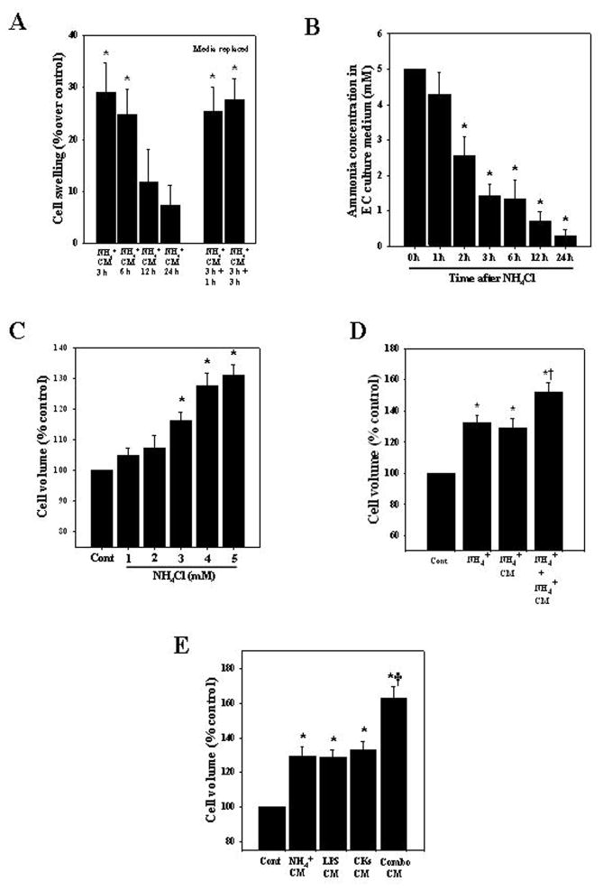
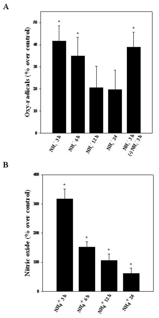
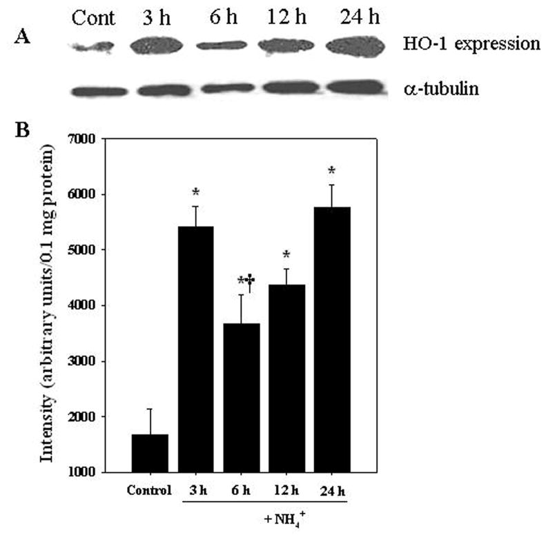
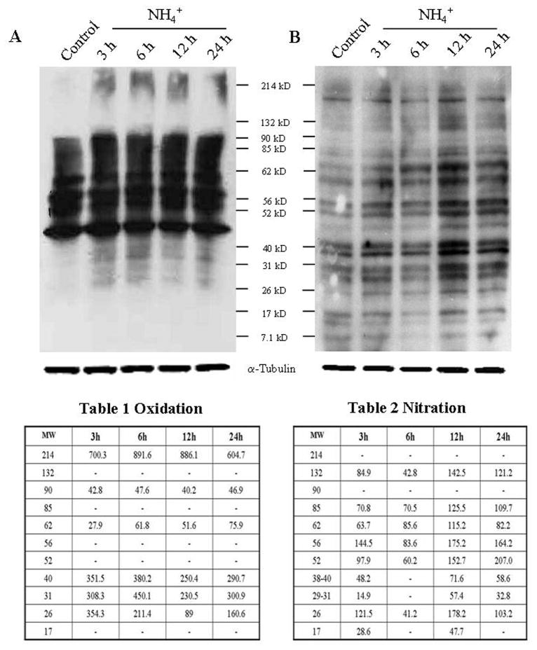
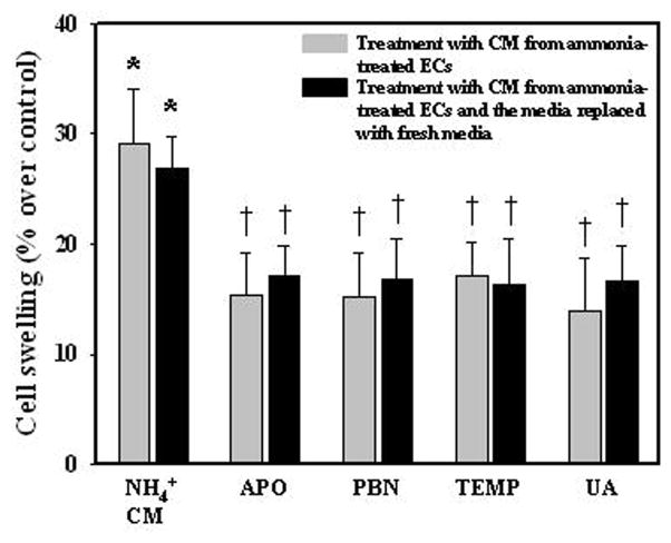
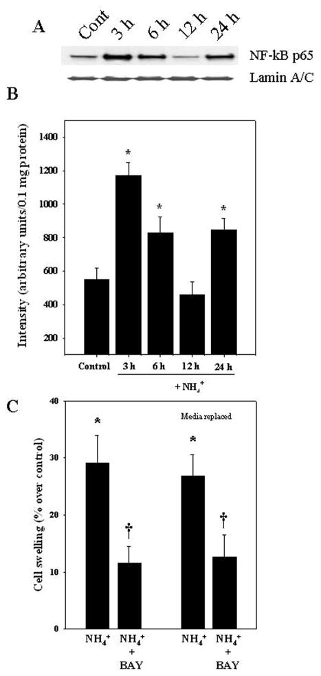
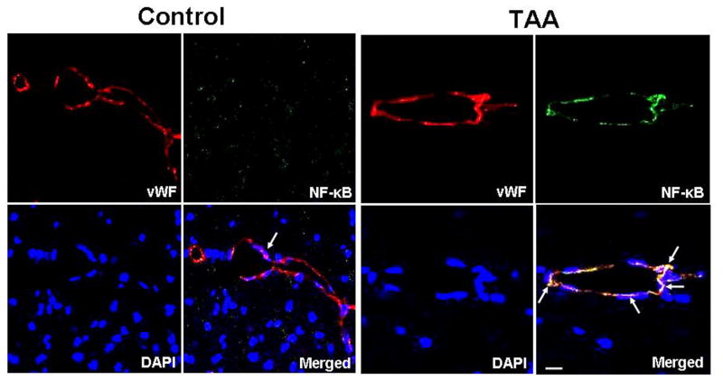
Brain edema is an important complication of acute hepatic encephalopathy (AHE), and astrocyte swelling is largely responsible for its development. Elevated blood and brain ammonia levels have been considered as major etiological factors in this edema. In addition to ammonia, recent studies have suggested that systemic infection, inflammation (and associated cytokines (CKs)), as well as endotoxin (lipopolysaccharide (LPS)) are also involved in AHE-associated brain edema. As endothelial cells (ECs) are the first resident brain cells exposed to blood-borne "noxious agents" (i.e., ammonia, CKs, LPS) that are present in AHE, these cells may be in a critical position to react to these agents and trigger a process resulting in astrocyte swelling/brain edema. We therefore examined the effect of conditioned media (CM) from ammonia, LPS and cytokine-treated cultured brain ECs on cell swelling in cultured astrocytes. CM from ammonia-treated ECs when added to astrocytes caused significant cell swelling, and such swelling was potentiated when astrocytes were exposed to CM from ECs treated with a combination of ammonia, LPS and CKs. We also found an additive effect when astrocytes were exposed to ammonia along with CM from ammonia-treated ECs. Additionally, ECs treated with ammonia showed a significant increase in the production of oxy-radicals, nitric oxide (NO), as well as evidence of oxidative/nitrative stress and activation of the transcription factor nuclear factor kappa B (NF-κB). CM derived from ECs treated with ammonia, along with antioxidants (AOs) or the NF-κB inhibitor BAY 11-7082, when added to astrocytes resulted in a significant reduction in cell swelling, as compared to the effect of CM from ECs-treated only with ammonia. We also identified increased nuclear NF-κB expression in rat brain cortical ECs in the thioacetamide (TAA) model of AHE. These studies suggest that ECs significantly contribute to the astrocyte swelling/brain edema in AHE, likely as a consequence of oxidative/nitrative stress and activation of NF-κB.


Figures

Figure 1
Figure 1
Cell swelling in cultured astrocytes after exposure to conditioned media (CM) from ammonia-treated endothelial cells (ECs). A. Cultured brain ECs were exposed to 5 mM NH4Cl for different time periods (3–24 h), the CM was added to astrocyte cultures for 24 h, and cell volume was determined. To exclude a potential role of any retained ammonia in the CM that might have contributed to the astrocyte swelling, ECs were treated with ammonia for 3 h, after which the ammonia-containing media was completely removed, and fresh media added to the ECs. At 1 and 3 h later the conditioned media from ECs was added to astrocytes and cell volume determined 24 h later (5th and 6th bars, media replaced). B. Ammonia concentration at different time points after ECs were treated with 5 mM NH4Cl. C. Time course of cell volume in ammonia-treated astrocyte cultures. D. Direct exposure of astrocyte cultures to ammonia, or to CM derived from ammonia-treated ECs (3 h) showed a significant increase in cell swelling, while a combination of these two treatments displayed a potentiation in cell swelling. E. ECs were treated with ammonia, LPS or with a mixture of cytokines (IL-1, IL-6, TNF-α, IFN-γ), alone or in combination (combo). CM from ammonia, LPS or mixture of cytokine-treated ECs when added to astrocytes caused a significant increase in astrocyte swelling, while CM from ECs treated with a combination of these agents resulted in a potentiation of astrocyte swelling. Data were subjected to ANOVA (n=7 for A; n=5 for B; n=4 for C; n=6 for D; n=5 for E). *p<0.05 vs. control. †p<0.05 vs. ammonia alone or with CM from ammonia-treated ECs in D; †p<0.05 versus ammonia, LPS or cytokines in E. Error bars, mean ± S.E. NH4+, ammonium chloride; LPS, lipopolysaccharide; CKs, a mixture of cytokines.
Figure 2
Figure 2
Time-dependent changes in free radical production in ECs following treatment with ammonia. A. Cultured ECs were exposed to ammonia (5 mM) for different time periods (3, 6, 12 and 24 h) and levels of free radicals in the culture medium were measured. A significant increase in oxy-radicals was observed at 3 and 6 h. Additionally, to establish whether free radicals (which might contribute to astrocyte swelling) can be identified after replacing the ammonia-containing media with fresh culture media, we measured free radicals in the freshly added cell culture media after 3 h. Free radicals were identified in the freshly added culture media (5th bar). B. A significant increase in nitric oxide was observed after ammonia treatment at all time points, with a peak at 3 h. Data were subjected to ANOVA (n=4). *p<0.05 vs. control. Error bars, mean ± S.E.
Figure 3
Figure 3
Heme oxygenase-1 (HO-1) protein expression in ECs following ammonia treatement. A. Representative western blots show a significant increase in HO-1 protein level when endothelial cultures were exposed to 5 mM NH4Cl. B. Quantification of NH4Cl-induced changes in HO-1 protein expression. HO-1 levels were normalized against α-tubulin. Data were subjected to ANOVA (n=5). *p<0.05 vs. control. †p<0.05 versus 3 and 24 h. Error bars, mean ± S.E.
Figure 4
Figure 4
Protein oxidation/nitration in ammonia-treated ECs. A. Oxidized proteins were detected by Western blot analysis with 2-DNPH. Proteins ranging in molecular weight from 26–40 kD as well as 62, 90 and 214 kD were found to be oxidized at all time points. Table 1, quantification of NH4Cl-induced changes in oxidized protein in ECs. B. Protein tyrosine nitration was also detected by Western blot analysis with an antibody raised against 3-nitrotyrosine. Proteins of molecular weight 26, 52–85 kD and 132 kD were found to be nitrated at all time points. Proteins of molecular weight 17 and 29–40 were nitrated at 3, 12 and 24 h after ammonia exposure (n=4). Table 2, quantification of NH4Cl-induced changes in nitrated protein in ECs. Oxidized and nitrated protein levels were normalized against α-tubulin.
Figure 5
Figure 5
Effect of CM from ECs treated with ammonia, in the presence or absence of antioxidants on astrocyte swelling. Astrocytes were exposed to CM derived from ammonia or ammonia plus antioxidants-treated ECs for 3 h. Ammonia plus antioxidant-treated ECs displayed a lesser degree of cell swelling as compared to astrocytes exposed to CM from only ammonia-treated ECs (gray bars). To rule-out the possibility that AOs in endothelial CM may have directly influenced the astrocyte swelling, ECs were treated with ammonia plus AOs for 3 h, after which the ammonia- and AO-containing media was removed, and fresh media added to the ECs. After 3 h the conditioned media from ECs was added to astrocyte cultures and cell volume determined 24 h later. This procedure also resulted in a comparable decrease in cell swelling (dark bars). Data were subjected to ANOVA (n=5 for gray bars and n=4 for dark bars). *p<0.05 vs. control. †p<0.05 vs. ammonia. Error bars, mean ± S.E. PBN, N-tert-butyl-α-phenylnitrone; APO, apocynin; TEMP, Tempol; UA, uric acid.
Figure 6
Figure 6
Nuclear translocation (activation) of NF-κ B in ammonia-treated ECs. Increased NF-κ B was identified in the nuclear fraction of cultured ECs that were treated with 5 mM NH4Cl. A. Western blots of NF-κ B (p65) in nuclear fraction. B. Quantification of Western blots. Ammonia caused an increase in nuclear NF-κ B at 3, 6, and 24 h, but had no effect at 12 h. NF-κ B content was normalized against lamin A/C. C. ECs were treated with BAY 11-7082, an inhibitor of NF-κ B, along with ammonia and the CM was added to astrocytes. Such treatment resulted in a lesser degree of cell swelling as compared to CM from ECs treated with ammonia alone. To exclude a direct effect of BAY 11-7082 on astrocyte swelling, ECs were treated with ammonia plus BAY 11-7082 for 3 h, after which the ammonia- and BAY 11-7082-containing media was removed, and fresh media added to the ECs. After 3 h the conditioned media from ECs was added to astrocyte cultures and cell volume determined 24 h later. This procedure also resulted in a comparable decrease in cell swelling (media replaced). ANOVA (n=4) for NF-κ B expression and n=5 for cell swelling studies. *p<0.05 versus control. †p<0.05 vs. ammonia. Error bars, mean ± S.E. BAY, BAY 11-7082.
Figure 7
Figure 7
Activation of NF-κ B in endothelial cells from cerebral cortex of rats with acute hepatic encephalopathy following the administration of the hepatotoxin thioacetamide (TAA) for 3 days. Control (normal) brain: von Willebrand Factor (vWF, red, endothelial cells), NF-κ B (green), and DAPI (blue, nuclei). The co-localization (merged) of NF-κ B, von Willebrand factor and DAPI illustrates that NF-κ B is not present in nuclei of von Willebrand factor-positive cells (arrows). TAA-treated rat brain: note the co-localization of NF-κ B and DAPI in vWF-positive cells, indicating nuclear localization (activation) of NF-κ B in endothelial cells (arrows). Scale bar =15 μm.

References

    1. Abbott NJ, Patabendige AA, Dolman DE, Yusof SR, Begley DJ. Structure and function of the blood-brain barrier. Neurobiol Dis. 2010;37:13–25. - PubMed
    1. Abbott NJ, Rönnbäck L, Hansson E. Astrocyte-endothelial interactions at the blood-brain barrier. Nat Rev Neurosci. 2006;7:41–53. - PubMed
    1. Albrecht J, Jones EA. Hepatic encephalopathy: molecular mechanisms underlying the clinical syndrome. J Neurol Sci. 1999;170:138–146. - PubMed
    1. Baldwin AS., Jr The NF-kappa B and I kappa B proteins: new discoveries and insights. Annu Rev Immunol. 1996;14:649–683. - PubMed
    1. Banks WA, Kastin AJ, Broadwell RD. Passage of cytokines across the blood-brain barrier. Neuroimmunomodulation. 1995;2:241–248. - PubMed

Publication types