. 2012 Aug 1;368(1):28-43.
doi: 10.1016/j.ydbio.2012.05.010. Epub 2012 May 17.

The FGF8-related signals Pyramus and Thisbe promote pathfinding, substrate adhesion, and survival of migrating longitudinal gut muscle founder cells

Affiliations

The FGF8-related signals Pyramus and Thisbe promote pathfinding, substrate adhesion, and survival of migrating longitudinal gut muscle founder cells

Ingolf Reim et al. Dev Biol. .

Abstract

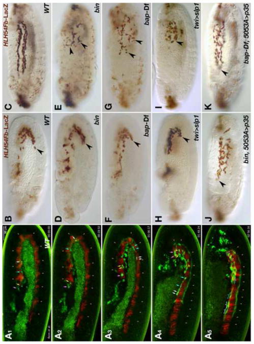
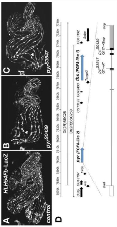
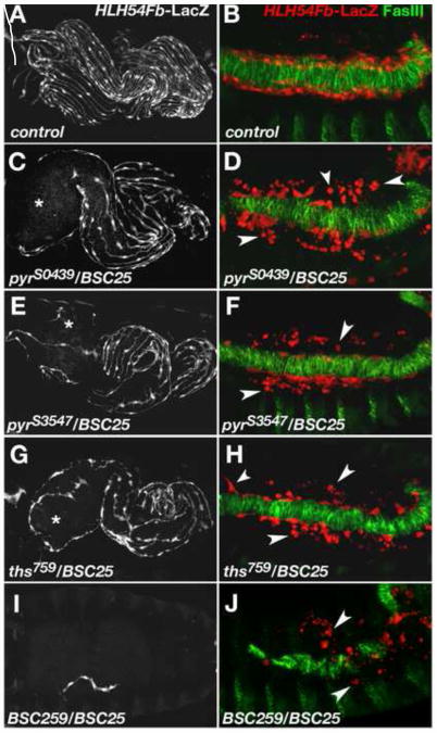
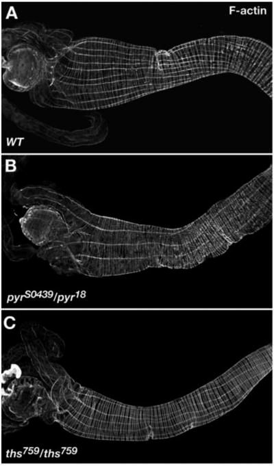
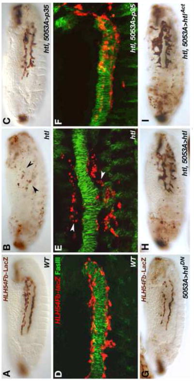
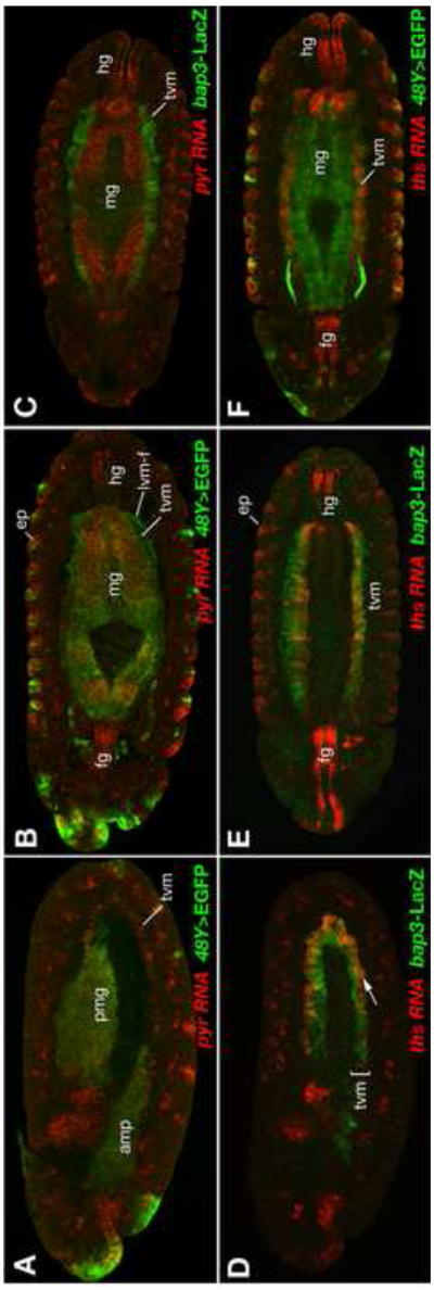
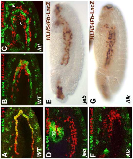
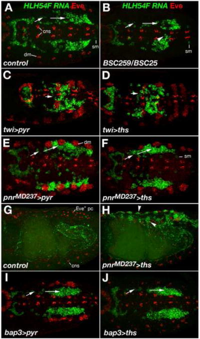
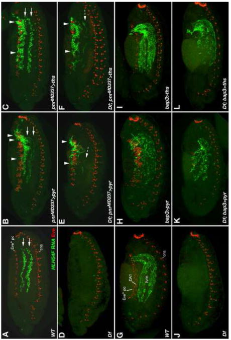
Fibroblast growth factors (FGFs) frequently fulfill prominent roles in the regulation of cell migration in various contexts. In Drosophila, the FGF8-like ligands Pyramus (Pyr) and Thisbe (Ths), which signal through their receptor Heartless (Htl), are known to regulate early mesodermal cell migration after gastrulation as well as glial cell migration during eye development. Herein, we show that Pyr and Ths also exert key roles during the long-distance migration of a specific sub-population of mesodermal cells that migrate from the caudal visceral mesoderm within stereotypic bilateral paths along the trunk visceral mesoderm toward the anterior. These cells constitute the founder myoblasts of the longitudinal midgut muscles. In a forward genetic screen for regulators of this morphogenetic process we identified loss of function alleles for pyr. We show that pyr and ths are expressed along the paths of migration in the trunk visceral mesoderm and endoderm and act largely redundantly to help guide the founder myoblasts reliably onto and along their substrate of migration. Ectopically-provided Pyr and Ths signals can efficiently re-rout the migrating cells, both in the presence and absence of endogenous signals. Our data indicate that the guidance functions of these FGFs must act in concert with other important attractive or adhesive activities of the trunk visceral mesoderm. Apart from their guidance functions, the Pyr and Ths signals play an obligatory role for the survival of the migrating cells. Without these signals, essentially all of these cells enter cell death and detach from the migration substrate during early migration. We present experiments that allowed us to dissect the roles of these FGFs as guidance cues versus trophic activities during the migration of the longitudinal visceral muscle founders.


Figures

Fig. 1
Fig. 1. Normal migration of longitudinal visceral muscle founders in the wild type and disrupted migration and survival in the absence of trunk visceral mesoderm
(A) Still images of wild type embryo from Movie 1 at five different time points (A1: 0 min; A2: 29 min, A3: 1 h 6 min, A4: 1 h 48 min, A5: 2 h 56 min). The embryo carries HLH54F-GFP, which marks the CVM and migrating LVM founders (green), as well as bap3-RFP, which marks the trunk visceral mesoderm (red; parasegmental borders marked by arrow heads). Four migrating LVM progenitors and their daughter cells are marked by color-coded arrows and the syncytia formed from these by brackets. (B – K) Embryos carrying HLH54Fb-lacZ are stained with anti-β-galactosidase (β-gal) antibodies at stage 11 (B, D, F, H• and late stage 13 (C, E, G, I-K). Shown are lateral views with the anterior on the left. (B) Wild type (WT) stage 11 control embryo. LacZ-positive LVM founder cells have left the posterior tip of the extended germ band and migrate around the U-turn toward the anterior on either side of the embryo. (C) At the end of stage 13 the leading cells from the migration paths along the dorsal and ventral edges of the TVM have reached the anterior of the trunk. (D, E) show bin22 and (F, G) bap null (Df(3R)e-D7, tin-re28) mutant embryos at corresponding stages. Early migration of the CVM is largely unaffected in these mutants. At stage 13, the most anterior cells have failed to migrate beyond the point reached already during stage 11 (arrow heads) and are scattered within in the posterior third of the embryo. They are also reduced in number, and many of the remaining LacZ-positive cells feature abnormal cell shapes and have shrunken in size. Similar defects are observed upon ectopic activation of slp1 via twi-GAL4 (H, I), which blocks TVM formation. (J, K) If cell death is prevented by expression of the caspase inhibitor p35 within LVM founders, the cells survive and regain their ability to migrate, albeit less orderly, into the anterior trunk in bin or bap mutant backgrounds.
Fig. 2
Fig. 2. Disrupted formation of longitudinal visceral muscles in homozygous pyr EMS mutants
(A) HLH54Fb-lacZ wild type embryo with anti-β-gal staining to visualize longitudinal visceral muscle fibers spaced evenly around the midgut all along its length at stage 17. (B) Homozygous pyrS0439 EMS mutant embryo carrying HLH54Fb-lacZ and stained as in (A), which shows a partial depletion of longitudinal visceral muscle fibers in the anterior portion of the midgut (asterisk). (C) Similar defects are seen in an embryo homozygous for the pyrS3547 allele. Note the wider LVM fiber spacing as compared to (A). In addition, gut looping abnormalities connected to an incomplete anterior midgut constriction are seen to variable degrees in both alleles. (D) Map of the genomic region containing pyr and ths. Shown are gene models contained near the areas of overlap of the deficiencies used to eliminate both FGF8-like genes. The gene structure of pyr and positions of the mapped pyr mutations are depicted below. Several other amino acid sequence variations compared to the published sequence were found in both the mutagenized and unmutagenized strains: A shortened poly-asparagine stretch at position 260 (from 10 to 4 N′s), Q316K, A400V, A599V, and M614V. Exons are boxed and shaded in their coding areas. Black shading indicates the region corresponding to the FGF domain (Pfam HMM consensus).
Fig. 3
Fig. 3. Abnormal longitudinal visceral muscle development in mutants with reduced FGF ligand activity
Anti-β-gal staining of HLH54Fb-lacZ embryos to visualize longitudinal visceral muscle fibers at stage 17 (A, C, E, G, I) and of migrating LVM progenitors at the end of germ band retraction (B, D, F, H, J). In the latter panels the TVM is marked by anti-fasciclin III (FasIII) staining in green. (A, B) Wild type. (C) Embryo carrying the EMS-induced mutation pyrS0439 in trans to the pyr- and ths-deleting deficiency Df(2R)BSC25 with a strong depletion of LVM fibers especially around the anterior midgut (asterisk). (D) Late stage 12 embryo of the same genotype as in (C). Numerous LVM founders have lost contact with the TVM and many of them appear as small rounded dots (arrow heads). (E, F) pyrS3547/Df(2R)BSC25 mutant embryo showing essentially the same phenotype as embryos in (C, D). Very similar defects are also observed in ths759/Df(2R)BSC25 mutants (G, H). (I) Removal of pyr and ths using overlapping deficiencies Df(2R)BSC25 and Df(2R)BSC259 (“FGF8 null” genotype) leads to a complete loss of the LVM with rare occurrence of individual LVM fibers. (J) In a FGF8 null mutant, the number of LVM founders at late stage 12 is greatly diminished and almost all cells have lost contact with the TVM.
Fig. 4
Fig. 4. Larval gut muscle phenotypes of pyr and ths mutants
Guts of 3rd instar larvae stained tor F-actin in visceral muscles using fluorescently labeled phalloidin. Shown are anterior portions with the proventriculus and gastric caecae to the left. (A) Wild type. (B) Gut from pyrS0439/pyr18 larva showing a reduction in the number of LVM fibers as compared to (A). (C) Gut from homozygous ths759 mutant larva also showing a reduction of LVM fibers.
Fig. 5
Fig. 5. Requirements for the FGF receptor Htl in migrating longitudinal visceral muscle founders
Stage 13 embryos carrying HLH54Fb-lacZ stained with anti-β-gal and DAB (A-C, G-I•• and higher magnification views of anti-β-gal/anti-FasIII fluorescent double stainings (D-F). (A, D) Wild type embryos with normal migration of LVM founder cells, which are spindle-shaped and closely associated with the dorsal and ventral edges of the TVM. (B, E) In htl loss-of-function mutants LVM founders do not migrate into the anterior and acquire small, rounded shapes characteristic of dying cells (arrow heads). (C, F) Upon inhibition of apoptosis in LVM founders via 5053A-GAL4 driven expression of p35, a majority of LVM cells is attached to the TVM at any given time and the cells are able to migrate almost as far as in the wild type. (G) LVM founder-specific expression of a dominant-negative form of htl using 5053A-GAL4. The number of LVM cells is reduced, there are scattered cell fragments, and migration is incomplete. (H, I) htl mutants in which htl function is specifically restored in LVM founders using 5053A-GAL4 in combination with GAL4-inducible transgenes encoding either a wild type (H) or a constitutively active form (I) of Htl. In both cases significant rescue of LVM cell survival and migration is observed (compare to B). In comparison with the wild type (A), the distances of anterior migration are reduced and many cells take abnormal directions. Especially the expression of ligand-independent receptors causes cells to form clusters with aberrant extensions in vertical directions (I).
Fig. 6
Fig. 6. Expression of pyr and ths during LVM founder cell migration and gut formation
Expression of pyr and ths RNA (red, as indicated) detected by in situ hybridization in embryos carrying either 48Y-GAL4/UAS-EGFP (A, B, F) or bap3-lacZ (C, D, E). 48Y-GAL4 drives GFP expression (green, anti-GFP) mainly in the endoderm, at low levels in the migrating LVM founder cells (lvm-f) but not in the trunk visceral mesoderm (tvm), and in some epidermal areas (ep). bap3-lacZ (green, anti-β-gal) is expressed exclusively in the trunk visceral mesoderm (tvm). (A) Lateral view of late stage 11 embryo showing pyr expression in the GFP-labeled primordia of the anterior and posterior midgut (amg, pmg) and in the row of TVM founder cells (tvm). (B, C) Ventral views of inner sections of stage 13 embryos demonstrating pyr co-expression with EGFP in the forming midgut endoderm (mg), but not in the TVM. (D) Lateral view of stage 11 embryo showing ths expression in the GFP-labeled primordia of the TVM. Within the TVM, only the ventrally located founder cells express ths (arrow). (E, F) Ventral views of inner sections of stage 13 embryos demonstrating ths co-expression with LacZ in the TVM, but not in the midgut (mg). Both genes, pyr and ths, show expression in the foregut (fg) and hindgut (hg) and in the epidermis (ep).
Fig. 7
Fig. 7. LVM founder migration without ths and pyr expression in the visceral mesoderm
(A) Double staining of ths RNA(green) together with Org-1 protein (red) in a stage 11 embryo. At this stage Org-1 labels the founder cells of the circular TVM. Co-staining confirms specific expression of ths in these cells. (B-D, F) Stage 12 embryos carrying the LVM marker HLH54Fb-lacZ stained with anti-β-gal (red) and in situ hybridized for detection of ths RNA (green). (B) In the wild type, LVM founders (red) migrate close to the ths expressing TVM founders (green). (C) htl mutant in which LVM founders (red) begin to lose contact with the TVM. Some of the LacZ-labeled cells have become small and rounded (arrow heads). (D) Stage 12 jeb mutant embryo showing normal migration and survival of LVM founders in the absence of TVM-expressed ths. (hg: ths in hindgut anlage) (E) Late stage 13 jeb mutant embryo stained with anti-β-gal to detect migration of HLH54Fb-lacZ expressing LVM cells at stage 13. The distance of migration is almost normal. LVM tracks are slightly irregular (compare with control in Fig. 5A and with htl mutant in Fig. 5B). (F) Stage 12 Alk mutant embryo showing normal migration and survival of LVM founders in the absence of TVM-expressed ths and pyr. (G) Late stage 13 Alk mutant embryo stained with anti-β-gal and showing the same LVM migration behaviour as jeb mutant in (E).
Fig. 8
Fig. 8. Effects of Pyr and Ths on the direction of LVM founder migration
Wild type, mutant, and FGF over-expressing embryos were stained for HLH54F RNA (green) and Even-skipped (Eve) protein (red). In A-F, I, and J, dorsal views of germ band extended embryos are shown with caudal ends to the left and anterior directions toward the right. (A) Stage 11 wild type embryo; HLH54F expression (green) marks migrating LVM founders derived from the caudal visceral mesoderm and Eve (red) marks FGF-dependent precursors of specific pericardial and somatic muscle cells in the dorsal mesoderm (dm). Arrows indicate the lateral-anterior directions of LVM founder migration. HLH54F is also weakly expressed in one ventrolateral somatic muscle progenitor per segment (sm) and Eve also labels cells of the central nervous system (cns; in a lower focal plain than the visceral mesoderm but included in the Z-stack projections as a reference for the location of the ventral midline). (B) FGF8 null mutant stage 11 embryo. At this stage, overall migration is relatively normal but less symmetric, with some cells straying from the normal path and migrating toward the midline and/or the Z direction (arrow head). The number of migrating LVM founders is slightly reduced. There is no eve activation in the dorsal mesoderm. (C, D) Ectopic expression of either pyr (C) or ths (D) in the entire mesoderm via twi-GAL4 (SG24) prevents LVM founders from forming bilateral groups. Migration of individual cells appears to be undirected with only little net movement toward the anterior trunk. Mesodermal Eve clusters are enlarged. (E, F) Ectopic expression of either pyr (E) or ths (F) in the dorsal ectoderm via pnrMD237-GAL4 causes LVM founders to migrate further laterally as compared to cells of the same stage in normal embryos. LVM founders between the (enlarged) Eve clusters of the dorsal mesoderm extend processes toward the ectoderm. (G) Lateral view of stage 15 wild type embryo in which LVM fibers have aligned along the entire midgut. Eve expression is seen dorsally in Eve+ pericardial cells, weakly in somatic muscle DA1, and ventrally in the CNS. (H) Stage 15 embryo with pnrMD237-GAL4-driven ectopic expression of ths in the dorsal ectoderm. LVM fibers are seen along the entire midgut as in (G), but many LVM cells have been redirected toward dorsal areas underneath the ectoderm (arrow heads). (I, J) LVM founders migrate normally in stage 11 embryos when either pyr (I) or ths (J) are over-expressed in the TVM via bap3-GAL4 (compare to A).
Fig. 9
Fig. 9. Rescue and re-routing of LVM founder migration in FGF8 null mutants by forced ectopic expression of pyr and ths
Wild type and mutant embryos with and without GAL4-driven FGF expression were stained for HLH54F RNA (green) and Even-skipped (Eve) protein (red) as in Fig. 8. Shown are lateral views of either stage 13 (A-F) or stage 14 (G-L) embryos. (A) In the wild type LVM precursors spread across the entire trunk along a dorsal and a ventral track (on each side of the embryo). (B) pnrMD237-GAL4-driven ectopic expression of pyr in the dorsal ectoderm of an otherwise wild type embryo leads to clustering of LVM precursors near the dorsal ectoderm (arrow heads). Many cells still take the normal route of migration (arrows). Eve expression in the dorsal somatic/cardiogenic area is expanded. (C) pnrMD237-GAL4-driven ectopic expression of ths has similar effects as pyr, although more cells are able to migrate to the anterior underneath the dorsal ectoderm and Eve expansion is less severe. (D) FGF8 null mutant (Df = Df(2R)BSC25/Df(2R)BSC259) in which almost no LVM forms. Pericardial/dorsal somatic Eve precursors are also absent. (E, F) In the absence of an endogenous FGF8-like ligand source, pnrMD237-GAL4-driven ectopic expression of either pyr or ths allows survival and migration of LVM founders. Almost all LVM cells cluster near the dorsal ectoderm and very few cells take a near normal route of migration (dashed arrows). In FGF8 null mutants with forced ths expression (F) Eve precursors are rescued only in some segments, but more LVM cells are able to migrate to the anterior than in mutants with forced pyr expression (E). (G) Wild type stage 14 embryo with several rows of HLH54F-expressing syncytial LVM fibers being formed. Eve expression is seen dorsally in Eve+ pericardial cells, weakly in somatic muscle DA1, and ventrally in the CNS. (H, I) bap3-GAL4-driven expression of pyr (H) or ths (I) in the TVM has little or no effect on the migration of LVM precursors except for mild disruptions in the arrangement of LVM fibers upon pyr over-expression. TVM-derived Pyr, but not Ths, also causes a strong expansion of dorsal Eve expression. (J) Stage 14 FGF8 null mutant (Df(2R)BSC25/Df(2R)BSC259) without any LVM and dorsal Eve expression. (K, L) Forced expression of either pyr or ths from a bap3-GAL4 controlled transgene rescues survival and migration of LVM founders in Df(2R)BSC25/Df(2R)BSC259 embryos. LVM formation proceeds, with some arrangement defects seen when driving pyr (K) and relatively normally when driving ths (L). Specification of Eve progenitors in the dorsal mesoderm is rescued partially (with pyr; K) or not at all (with ths; L).

Similar articles

Cited by

References

    1. Azpiazu N, Frasch M. tinman and bagpipe: two homeo box genes that determine cell fates in the dorsal mesoderm of Drosophila. Genes Dev. 1993;7:1325–1340. - PubMed
    1. Baker R, Schubiger G. Autonomous and nonautonomous Notch functions for embryonic muscle and epidermis development in Drosophila. Development. 1996;122:617–626. - PubMed
    1. Beiman M, Shilo B, Volk T. Heartless, a Drosophila FGF receptor homolog, is essential for cell migration and establishment of several mesodermal lineages. Genes Dev. 1996;10:2993–3002. - PubMed
    1. Brückner K, Kockel L, Duchek P, Luque CM, Rorth P, Perrimon N. The PDGF/VEGF receptor controls blood cell survival in Drosophila. Dev Cell. 2004;7:73–84. - PubMed
    1. Cabernard C, Affolter M. Distinct roles for two receptor tyrosine kinases in epithelial branching morphogenesis in Drosophila. Dev Cell. 2005;9:831–842. - PubMed

Publication types

MeSH terms

LinkOut - more resources