

The prevalence and diagnostic utility of endoscopic features of eosinophilic esophagitis: a meta-analysis
- PMID: 22610003
- PMCID: PMC3424367
- DOI: 10.1016/j.cgh.2012.04.019
The prevalence and diagnostic utility of endoscopic features of eosinophilic esophagitis: a meta-analysis
Abstract
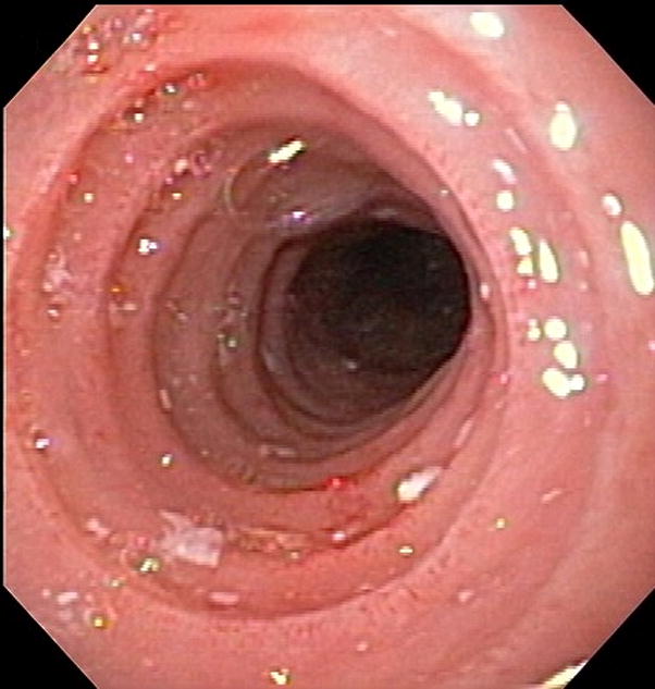
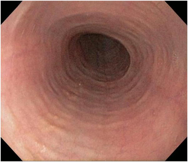
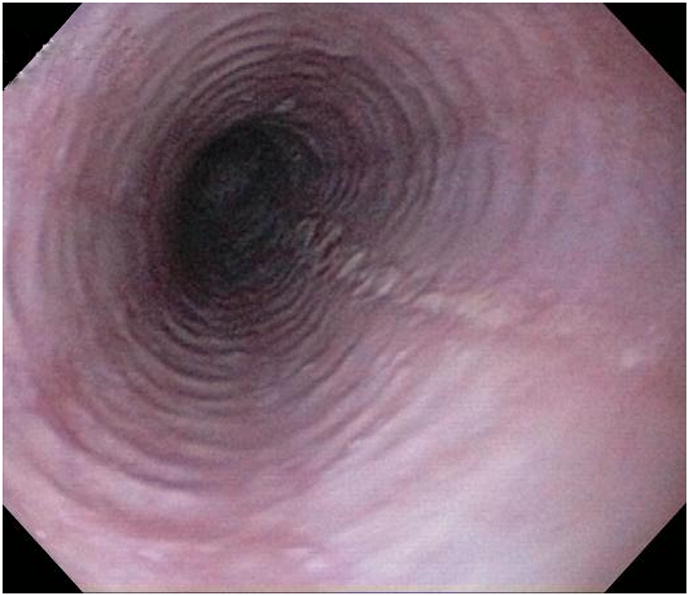
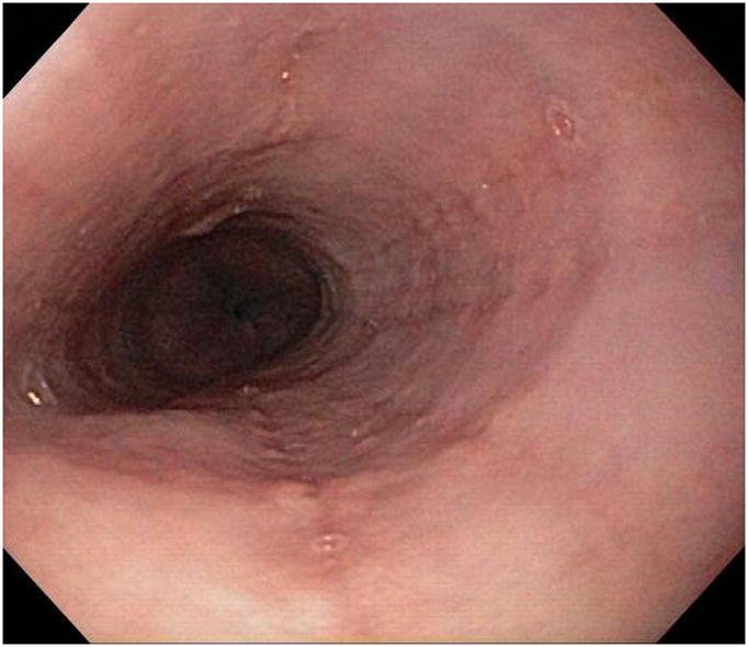
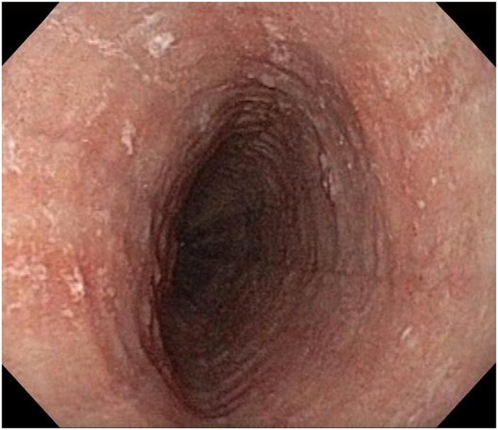
Background & aims: Endoscopic findings such as esophageal rings, strictures, narrow-caliber esophagus, linear furrows, white plaques, and pallor or decreased vasculature might indicate the presence of eosinophilic esophagitis (EoE). We aimed to determine the prevalence and diagnostic utility of endoscopic features of EoE.
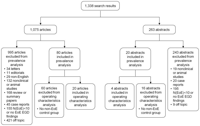
Methods: We conducted a systematic review and meta-analysis. PubMed, EMBASE, and gastrointestinal meeting abstracts were searched to identify studies that included more than 10 patients with EoE and reported endoscopic findings. Pooled prevalence, sensitivity, specificity, and predictive values were calculated using random- and mixed-effects models.
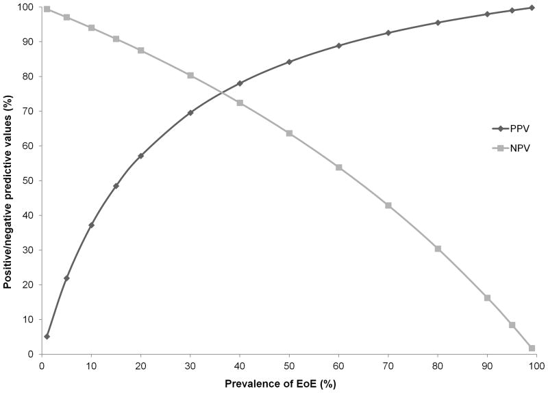
Results: The search yielded 100 articles and abstracts on 4678 patients with EoE and 2742 without (controls). In subjects with EoE, the overall pooled prevalence was as follows: esophageal rings, 44%; strictures, 21%; narrow-caliber esophagus, 9%; linear furrows, 48%; white plaques, 27%; and pallor/decreased vasculature, 41%. Substantial heterogeneity existed among studies. Results from endoscopy examinations were normal in 17% of patients, but this number decreased to 7% when the analysis was limited to prospective studies (P < .05). Overall levels of sensitivity were modest, ranging from 15% to 48%, whereas levels of specificity were greater, ranging from 90% to 95%. Positive predictive values ranged from 51% to 73% and negative predictive values ranged from 74% to 84%.
Conclusions: There is heterogeneity among studies in the reported prevalence of endoscopic findings in patients with EoE, but in prospective studies at least 1 abnormality was detected by endoscopy in 93% of patients. The operating characteristics of endoscopic findings alone are inadequate for diagnosis of EoE. Esophageal biopsy specimens should be obtained from all patients with clinical features of EoE, regardless of the endoscopic appearance of the esophagus.
Copyright © 2012 AGA Institute. Published by Elsevier Inc. All rights reserved.
Figures
References
-
- Landres RT, Kuster GG, Strum WB. Eosinophilic esophagitis in a patient with vigorous achalasia. Gastroenterology. 1978;74(6):1298–301. Epub 1978/06/01. - PubMed
-
- Furuta GT, Liacouras CA, Collins MH, Gupta SK, Justinich C, Putnam PE, et al. Eosinophilic esophagitis in children and adults: a systematic review and consensus recommendations for diagnosis and treatment. Gastroenterology. 2007;133(4):1342–63. Epub 2007/10/09. - PubMed
-
- Ferre-Ybarz L, Nevot Falco S, Plaza-Martin AM. Eosinophilic oesophagitis: clinical manifestations and treatment options. The role of the allergologist. Allergologia et immunopathologia. 2008;36(6):358–65. Epub 2009/01/20. - PubMed
-
- Sgouros SN, Bergele C, Mantides A. Eosinophilic esophagitis in adults: a systematic review. European journal of gastroenterology & hepatology. 2006;18(2):211–7. Epub 2006/01/06. - PubMed
Publication types
MeSH terms
Grants and funding
LinkOut - more resources
Full Text Sources
Other Literature Sources
Medical
