. 2012 Jun;15(6):862-70.
doi: 10.1038/nn.3109.

Hepatocyte growth factor mediates mesenchymal stem cell–induced recovery in multiple sclerosis models

Affiliations

Hepatocyte growth factor mediates mesenchymal stem cell–induced recovery in multiple sclerosis models

Lianhua Bai et al. Nat Neurosci. 2012 Jun.

Abstract

Mesenchymal stem cells (MSCs) have emerged as a potential therapy for a range of neural insults. In animal models of multiple sclerosis, an autoimmune disease that targets oligodendrocytes and myelin, treatment with human MSCs results in functional improvement that reflects both modulation of the immune response and myelin repair. Here we demonstrate that conditioned medium from human MSCs (MSC-CM) reduces functional deficits in mouse MOG35–55-induced experimental autoimmune encephalomyelitis (EAE) and promotes the development of oligodendrocytes and neurons. Functional assays identified hepatocyte growth factor (HGF) and its primary receptor cMet as critical in MSC-stimulated recovery in EAE, neural cell development and remyelination. Active MSC-CM contained HGF, and exogenously supplied HGF promoted recovery in EAE, whereas cMet and antibodies to HGF blocked the functional recovery mediated by HGF and MSC-CM. Systemic treatment with HGF markedly accelerated remyelination in lysolecithin-induced rat dorsal spinal cord lesions and in slice cultures. Together these data strongly implicate HGF in mediating MSC-stimulated functional recovery in animal models of multiple sclerosis.


Figures
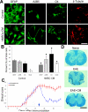
Figure 1
Figure 1
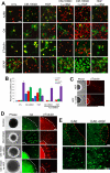
Conditioned growth medium from human MSCs biases the development of neurosphere derived cells towards oligodendrocytes and neurons and promotes functional recovery in MOG35–55 induced EAE. A. In the presence of human MSC-CM the proportion or GFAP+ astrocytes are reduced while the proportion of oligodendrocyte lineage cells and neurons is increased. B. Quantitation of relative number of distinct cell types in the presence and absence of MSC-CM. (Control vs. MSC-CM, A2B5 p=<0.01, O4 p=<0.05, β–tubulin p=<0.05, GFAP p=0.005) C. Treatment with MSC-CM (0.5mg) (arrow), but not PBS, at peak of disease after MOG35–55 immunization results in functional improvement in EAE. D. The functional improvement is correlated with a reduction in myelin loss and tissue damage seen with Luxol Fast Blue staining of spinal cord sections. Lesions are outlined in D. Data in B represent the mean±SEM of duplicate preparations taken from 3 independent experiments. Note there is both overlapping expression and non-labeled cells in these preparations. Bar = 50μm in A, 500μm in D.
Figure 2
Figure 2
The activity of MSC-CM to enhance functional recovery in EAE is dependent on a 1–100kD fraction. A. Treatment with MSC-CM100kD (0.5mg/animal n=11), but not control CM, enhances functional recovery in animals with EAE. B. Growth of neurosphere-derived cultures in CM100kD biases cell development in favor of oligodendrocytes and neurons compared to control CM. C. Treatment with CM100kD reduces proinflammatory cytokine expression by spinal cord derived mononuclear cells in EAE animals. Significantly reduced expression of IFNγ, IL-17, TNFα, IL-2 and IL-12p70 and increased expression of IL-10 and IL-4 were seen in animals treated with MSC-CM100kD. Results represent the mean±SEM from three independent experiments.
Figure 3
Figure 3
Human MSC-CM100kD contains HGF and HGF promotes functional and histological recovery in EAE. A. Western blot of 3 samples of active MSC-CM100kD show the presence of HGF. B. Treatment with HGF (1 injection every other day for a total of 3 over a 5 day period, n=10) results in functional improvement compared to controls. Both dosage and route of delivery influence the efficacy of HGF to modulate disease. Intravenous (IV) injections are more effective than intraperitoneal (IP) injections. Arrows indicate treatment initiation day. C. HGF improves tissue histology. Sections from animals following 100ng IV injections at 30 days after immunization with MOG35–55. The improvement in myelination is apparent by Luxol Fast Blue staining. The reduction in immune cell infiltrates is evident in H&E and anti-CD3 labeled sections and confirmed in Toluidine Blue stained 1μm sections. Lower panel are representative electron micrographs through spinal cord lesion areas in EAE control and HGF treated animals 17 days after initiation of treatment. D. Upper Analysis of myelin thickness vs. axon diameter in lesion areas of control and HGF treated animals demonstrates thicker myelin in HGF treated animals compared to controls. Lower: Comparison of the relative axons diameters in lesion areas of control EAE and HGF treated animals demonstrates a reduction in small diameter fibers and an increase in medium diameter fibers in HGF treated animals. Bars = 500μm in C LFB (top), 50μm in C LFB (2ndpanel), H&E, CD3, Tol Blue and 2μm in the electron micrographs.
Figure 4
Figure 4
Inhibition of HGF signaling with cMet antibodies negates the capacity of both HGF and MSC-CM to induce functional recovery and reverses EAE induced changes in cytokine expression. A. Two injections of function blocking cMet antibodies 12hrs apart, (green arrows) delivered 1 day prior to initiation of HFG treatment regime (red arrows) inhibits functional recovery. B. Treatment with cMet antibodies increases proinflammatory cytokine expression and reduces anti-inflammatory cytokine expression as shown by cytokine profile. C. ELISPOT analysis on spinal cord derived cells demonstrates that treatment with cMet antibodies increases the frequencies of TH-1/TH-17 cells compared to HGF treated animals. Similar data (D,E,F) was obtained following cMet inhibition of MSC-CM100kD stimulated recovery. Treatment of MSC-CM100kD with anti-HGF antibodies also blocked its capacity to enhance functional recovery in EAE and inhibited MSC-CM induced reduction in pro-inflammatory cytokine expression. The delay in disease onset reflects the use of a different preparation of MOG35–55 peptide and not anti-HGF treatment. The data are representative from triplicate studies and counts represent the mean±standard deviation from one of three experiments.
Figure 5
Figure 5
Inhibition of HGF signaling with cMet or anti-HGF antibodies blocks the ability of MSC-CM100kD and HGF to alter the development and migration of neural cells from neurospheres. A. The proportions of A2B5+, O4+, β tubulin + and GFAP+ cells are altered in the presence of MSC-CM100kD and HGF and reversed by cMet and anti-HGF. B. Quantification of different cell types in the presence and absence of MSC-CM100kD and HGF± cMet antibodies. Compared to controls the proportion of O4+ and β tubulin + cells is increased by MSC-CM and HGF (p=<0.05 for both) and blocked by cMet antibodies (p=<0.01 for both) C. Treatment with HGF stimulates migration of neuronal precursors from adult SVZ derived neurospheres of EAE animals and is blocked by cMet antibodies. D. Treatment with MSC-CM100kD stimulates migration of OPCs and neuronal precursors from adult EAE SVZ derived neurospheres and is blocked by cMet and anti-HGF. E. The mobilization of PLP+ OPCs into EAE lesions is enhanced in animals treated with HGF. The number of EGFP-PLP cells which populate lesions increase following HGF treatment.. Bars = 20μm in A and 100μm in C and D. and 50μm in E. Data in B represent the mean±SD of the proportion of individual cell types taken from 5 random fields from at least 2 independent experiments. Lesion areas are outlined in E. Note that in B there is both unlabeled and overlap of antigen expression on individual cells.
Figure 6
Figure 6
Systemic HGF treatment stimulates remyelination of rat spinal cord LPC lesions. A,B Treatment with 0.8mg/Kg HGF on days 5,9 and 11 post lesion results in 30% smaller demyelinated lesions on 14dpl as shown by Luxol Fast Blue. Lesion volumes were calculated from serial sections through the lesion based on the equation for an elliptical cone (V=pi/3*a*b*h) and location of the lesion midpoint. C–H Immunohistochemical labeling of frozen sections demonstrated an increase in MBP labeling (C,D), increase in NG2+ cells (E,F) and a decrease in GFAP expression (G,H) in lesion areas from HGF treated animals at 14 days post lesion. I–J 1μm Toluidine Blue sections show extensive remyelination at 14 dpl in HGF treated animals but not in controls and ultrastructural analyses showed that axons of different caliber had myelin sheaths of different thicknesses indicative of ongoing repair in HGF (K,L,M) but not control lesions (K). N G ratios showing thin myelin sheaths in HGF treated animals.. Bars = A–F =100μm, G,H=50μm, I,J =5μm, K= 1μm, L=2μm, M= 0.5μm.

References

    1. Prineas JW. The neuropathology of multiple sclerosis. In: Koestier JC, editor. Handbook of clinical neurology. Elsevier; Amsterdam: 1985.
    1. Trapp BD, et al. Axonal transection in the lesions of multiple sclerosis. New England Journal of Medicine. 1998;338:278–285. - PubMed
    1. Wolswijk G, Noble M. Identification of an adult-specific glial progenitor cell. Development. 1989;105:387–400. - PubMed
    1. Reynolds BA, Weiss S. Generation of neurons and astrocytes from isolated cells of the adult mammalian central nervous system. Science. 1992;255:1707–1710. - PubMed
    1. Ben-Hur T, Goldman SA. Prospects of cell therapy for disorders of myelin. Annals of the New York Academy of Sciences. 2008;1142:218–249. - PubMed

Publication types

MeSH terms

Substances