Review
. 2012 Sep;1823(9):1468-83.
doi: 10.1016/j.bbamcr.2012.05.010. Epub 2012 May 17.

Mammalian iron metabolism and its control by iron regulatory proteins

Affiliations
Review

Mammalian iron metabolism and its control by iron regulatory proteins

Cole P Anderson et al. Biochim Biophys Acta. 2012 Sep.

Abstract

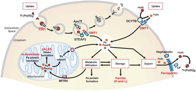
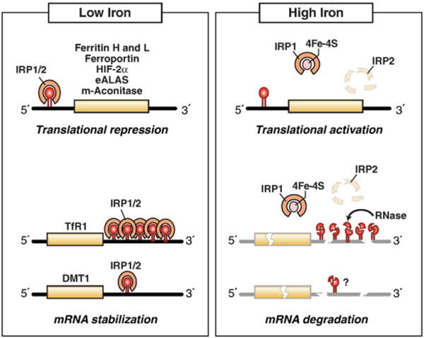
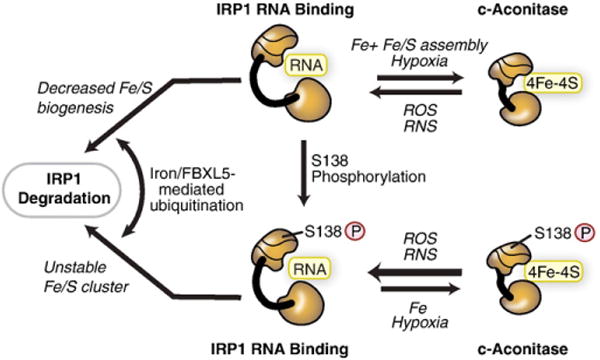
Cellular iron homeostasis is maintained by iron regulatory proteins 1 and 2 (IRP1 and IRP2). IRPs bind to iron-responsive elements (IREs) located in the untranslated regions of mRNAs encoding protein involved in iron uptake, storage, utilization and export. Over the past decade, significant progress has been made in understanding how IRPs are regulated by iron-dependent and iron-independent mechanisms and the pathological consequences of IRP2 deficiency in mice. The identification of novel IREs involved in diverse cellular pathways has revealed that the IRP-IRE network extends to processes other than iron homeostasis. A mechanistic understanding of IRP regulation will likely yield important insights into the basis of disorders of iron metabolism. This article is part of a Special Issue entitled: Cell Biology of Metals.


Figures

Fig. 1
Fig. 1
Control of mammalian cellular iron homeostasis by the IRE–IRP regulatory network. A generic mammalian cell depicting roles of proteins encoded by IRE-containing mRNAs (red lettering). Transferrin bound to two iron atoms (Tf-[Fe(III)2) binds to TfR1 on the cell surface where the Tf-[Fe(III)2–TfR1 complex is endocytosed. Acidification of the endosome causes the release of Fe(III) (red balls) from Tf where it is reduced to Fe(II) (orange balls) by the STEAP3 oxidoreductase before export by DMT1 (divalent metal transporter 1). ApoTf/TfR1 complex is returned to the cell surface where it dissociates and initiates another round of iron uptake. Tf-bound iron is taken up by most cells, but it is especially important in erythroid precursors where it is the primary source of iron for heme synthesis. DMT1 is also localized on the apical membrane of duodenal enterocytes where it transports Fe(II) after reduction by membrane reductases, such as DCYTB. Iron taken up by either TfR1 or DMT1 enters a cytosolic free labile iron pool thought to consist of Fe(II) bound to small molecular weight molecules. IRPs sense iron in this pool and regulate the translation of 5′ IRE-containing mRNAs (H-ferritin and L-ferritin, eALAS (erythroid aminolevulinate synthase), mitochondrial (m)-aconitase and ferroportin) or the stability of 3′ IRE-containing mRNAs (TfR1 and DMT1). eALAS serves as the rate-limiting enzyme in heme synthesis in erythroid precursors. Mitochondrial aconitase is an enzyme in the TCA cycle that requires a [4Fe–4S] cluster for activity. Iron that is not utilized or stored in ferritin is exported by ferroportin. Export of iron from cells is coupled to the oxidation of iron by membrane-bound hephaestin or serum multicopper oxidase ceruloplasmin (CP). Modified from Wallander et al. [4].
Fig. 2
Fig. 2
IRPs regulate translation and stability of IRE-containing mRNAs. IRPs bind to IREs located in either the 5′ or 3′ untranslated regions of specific mRNAs. When iron is limited, IRPs bind with high affinity to 5′ IRE mRNAs and repress translation, and to the five 3′ IREs in TfR1 mRNA and to the single IRE in DMT1 mRNA and stabilize these mRNAs. When iron is abundant, IRPs do not bind IREs, resulting in the translation of 5′ IRE-containing mRNAs and degradation of TfR1 mRNA. Iron mediates the conversion of the IRP1 RNA binding form into the [4Fe–4S] cluster c-aconitase form and the ubiquitination and targeted proteasomal degradation IRP2 by FBXL5 E3 ligase. IRE-containing mRNAs indicated are those that have been shown to be functional in vivo. Modified from Wallander et al. [4].
Fig. 3
Fig. 3
Iron-dependent and iron-independent mechanisms for regulating IRP1. IRP1 can be regulated through mechanisms dependent or independent of the [4Fe–4S] cluster. In the cluster-dependent pathway, IRP1 has dual roles either as a high affinity IRE-binding protein when devoid of the [4Fe–4S] cluster or as a c-aconitase when the [4Fe–4S] cluster is assembled. The c-aconitase form does not bind RNA. The [4Fe–4S] cluster is accessible to low molecular cluster perturbants, including reactive oxygen species (ROS), such as O2, H2O2 or reactive nitrogen species (RNS), such as NO or ONOO. Hypoxia stabilizes the c-aconitase form by reducing the level of oxygen or ROS. IRP1 can be phosphorylated at S138. IRP1 phosphomimetic mutants indicate that the Fe–S cluster can be assembled and the protein exhibits aconitase activity; however, the Fe–S cluster is more sensitive to disruption by oxygen and hydrogen peroxide. Iron stimulates the FBXL5-mediated degradation of IRP1 S138 phosphomimetic mutants and non-phosphorylated IRP1 when Fe–S cluster biogenesis is impaired. Modified from Wallander et al. [4].
Fig. 4
Fig. 4
Altered affinity of IRP1 for mutant and natural IREs. A) The affinity of interaction of IRP1 for six vertebrate 5′ IREs as determined by electrophoretic mobility shift assays (EMSA) [108]. The KD for these IREs varied over a 9-fold range. B) Mutations in the human L-ferritin IRE were identified in seven patients with hereditary hyperferritinemia–cataract syndrome. The affinity of interaction of IRP1 with mutant human L-ferritin IREs was determined by EMSA and related to serum ferritin values reported for each patient on different occasions (open circles) [113]. Maximal serum ferritin for each patient (closed circles). Results on the x-axis are expressed as Krel which is KD,mutant/KD,wildtype and higher values for Krel indicate lower binding affinity of the mutant IRE with IRP1. Note the steeper slope of the relationship between serum ferritin and Krel over the range 1 to 10 for Krel. A similar observation was made for IRP2 [113].
Fig. 5
Fig. 5
Comparison of the proposed secondary structure of IREs. The key structural elements required for recognition of the IRE by IRP1 include the unpaired C (C8) in the IRE stem and the A16G17U18 nucleotides of the pseudotriloop. The canonical IRE secondary structure is represented by one of the five TfR1 IREs (TfR1 B), where the RNA helix is interrupted by only the unpaired C8. The L-ferritin IRE secondary structure showing the conserved unpaired U at position 6 (arrow). This additional unpaired nucleotide has been observed in the NMR of the ferritin IRE and the crystal structure of IRP1 bound to the ferritin IRE [124,130,133]. Proposed secondary structures of DMT1 and HIF-2α IREs. Note the additional unpaired nucleotides (arrows). IRE residue numbering is according to Walden et al. [130].
Fig. 6
Fig. 6
Crystal structure of c-aconitase and the IRP1:IRE complex. The crystal structures of c-aconitase [132] and the IRP1:IRE complex [121,130] are shown. A) Cytosolic aconitase structure showing domain 1 (yellow), domain 2 (green), domain 3 (blue) and domain 4 (red) with the [4Fe–4S] cluster in the center (orange balls). B) IRP1:IRE complex structure shown with domains 1 to 4 as in (A). The ferritin IRE helix (purple) is shown with the two major contact sites of C8 (left) and the A16G17U18 bases of the pseudotriloop shown (right) as enlarged balls.
Fig. 7
Fig. 7
Mechanisms for iron-dependent and iron-independent IRP2 regulation. Iron-dependent degradation: when iron and oxygen are high, the FBXL5 hemerythrin-domain binds iron and leads to a conformational change that increases FBXL5 stability. FBXL5 binds to IRP2 and FBXL5-IRP2 associates with SKP1–CUL1 complex, catalyzing the ubiquitination and proteasomal degradation of IRP2. When iron or oxygen is limited, FBXL5 is destabilized and targeted for degradation by an unidentified E3 ligase. IRP2 iron-independent pathway: IRP2 is phosphorylated by cyclin-dependent kinase 1(CDK1/cyclinB1) during G2/M and dephosphorylated by CDC14A at the end of mitosis. S157 phosphorylation is independent of iron and is associated with reduced RNA binding activity. Non-phosphorylated and phosphorylated IRP2 are subjected to FBXL5 iron-mediated degradation. Modified from Wallander et al. [4].

References

    1. Fleming RE, Ponka P. Iron overload in human disease. N Engl J Med. 2012;366:348–359. - PubMed
    1. Madsen E, Gitlin JD. Copper and iron disorders of the brain. Annu Rev Neurosci. 2007;30:317–337. - PubMed
    1. Simmons DA, Casale M, Alcon B, Pham N, Narayan N, Lynch G. Ferritin accumulation in dystrophic microglia is an early event in the development of Huntington’s disease. Glia. 2007;55:1074–1084. - PubMed
    1. Wallander ML, Leibold EA, Eisenstein RS. Molecular control of vertebrate iron homeostasis by iron regulatory proteins. Biochim Biophys Acta. 2006;1763:668–689. - PMC - PubMed
    1. Rouault TA. The role of iron regulatory proteins in mammalian iron homeostasis and disease. Nat Chem Biol. 2006;2:406–414. - PubMed

Publication types