. 2012 Jul 13;287(29):24505-15.
doi: 10.1074/jbc.M112.347104. Epub 2012 May 18.

Arrestin scaffolds NHERF1 to the P2Y12 receptor to regulate receptor internalization

Affiliations

Arrestin scaffolds NHERF1 to the P2Y12 receptor to regulate receptor internalization

Shaista P Nisar et al. J Biol Chem. .

Abstract

We have recently shown in a patient with mild bleeding that the PDZ-binding motif of the platelet G protein-coupled P2Y(12) receptor (P2Y(12)R) is required for effective receptor traffic in human platelets. In this study we show for the first time that the PDZ motif-binding protein NHERF1 exerts a major role in potentiating G protein-coupled receptor (GPCR) internalization. NHERF1 interacts with the C-tail of the P2Y(12)R and unlike many other GPCRs, NHERF1 interaction is required for effective P2Y(12)R internalization. In vitro and prior to agonist stimulation P2Y(12)R/NHERF1 interaction requires the intact PDZ binding motif of this receptor. Interestingly on receptor stimulation NHERF1 no longer interacts directly with the receptor but instead binds to the receptor via the endocytic scaffolding protein arrestin. These findings suggest a novel model by which arrestin can serve as an adaptor to promote NHERF1 interaction with a GPCR to facilitate effective NHERF1-dependent receptor internalization.


Figures
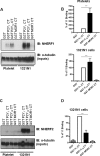
FIGURE 1.
FIGURE 1.
In vitro association of NHERF proteins isoforms with the C-tail of the P2Y1 and P2Y12R. A and C, human platelet (first to fourth lanes) or 1321N1 (fifth to eighth lanes) cell lysates were incubated with GST fusion proteins containing the P2Y1, P2Y12, or MOR1 receptor cytoplasmic tails (CT). Samples were resolved by SDS-PAGE and immunoblotted (IB) for NHERF1 (A) or NHERF2 (C). Inputs were immunoblotted for α-tubulin to show equal protein across samples. Blots shown are representative of five independent experiments. Multiple experiments were quantified by densitometry (B) shows quantified NHERF1 binding from platelets and 1321N1 cell lysates, respectively, **, p < 0.05 comparing level of P2Y1/NHERF1 to level of P2Y12/NHERF1 interaction (Mann-Whitney U test) (D) shows quantified NHERF2 binding from 1321N1 cell lysates. **, p < 0.05 comparing level of P2Y1/NHERF2 to level of P2Y12/NHERF2 interaction (Mann Whitney U test) (data are represented as mean ± S.E. (n = 5) in B.
FIGURE 2.
FIGURE 2.
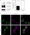
Agonist-dependent association of NHERF1 with full-length P2Y12R. A, 1321N1 cells stably expressing HA-tagged P2Y12R were incubated in the absence or presence ADP (10 μm, 5 min). Cells were lysed and receptors were immunoprecipitated using an HA antibody. Samples were resolved by SDS-PAGE and immunoblotted for associated NHERF1 (top panel) and reprobed with an anti-HA antibody to show total receptor immunoprecipitated (middle panel). Inputs were immunoblotted for NHERF1 (bottom panel) to show equal protein concentrations across samples. Blots shown are representative of three independent experiments. B, quantification of co-immunoprecipitated NHERF1 with P2Y12R (mean ± S.E., n = 3). **, p < 0.05 comparing level of basal P2Y12/NHERF1 interaction versus ADP-stimulated P2Y12/NHERF1 interaction (Mann-Whitney U test). C, agonist-dependent co-localization of P2Y12R and NHERF1 in human platelets. Human platelets were isolated and surface P2Y12 receptors were labeled with an anti-P2Y12 antibody (green). Platelets were stimulated in the absence (top panel) or presence (lower panel) of ADP (10 μm, 5 min). Endogenous NHERF1 was stained using an anti-NHERF1 antibody (purple). Co-localization is shown in the merged images (white). Images shown are representative of three independent experiments. Scale bar represents 10 μm.
FIGURE 3.
FIGURE 3.
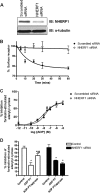
P2Y12R internalization is NHERF1-dependent. 1321N1 cells stably expressing HA-P2Y12R were treated with control or NHERF1 siRNA. A, 1321N1 cell lysates derived from control or NHERF1 siRNA-treated cells were subjected to NHERF1 immunoblotting to show NHERF1 protein knockdown. Membranes were re-probed with an anti-α-tubulin antibody to ensure equal protein loading. B, control or NHERF1 siRNA-treated cells were challenged with ADP (10 μm ADP; 0–60 min). Changes in cell surface receptor expression were evaluated by ELISA. C, P2Y12R activity in control and NHERF1 siRNA-treated cells. ADP-dependent inhibition of forskolin (1 μm; 10 min)-stimulated adenylyl cyclase activity was evaluated in control and NHERF1 siRNA-treated cells at a range of ADP concentrations. D, P2Y12R desensitization and subsequent desensitization were assessed by comparing agonist (ADP; 10 μm)-dependent inhibition of forskolin (1 μm; 10 min)-stimulated adenylyl cyclase activity before (control) and after pretreatment with either ADP alone (10 nm; 15 min; ADP-PT on graph) or after subsequent removal of desensitizing ADP with apyrase (0.2 units/ml; 15 min; ADP PT/Apyrase on graph). Data represent mean ± S.E. of 4 independent experiments. *, p < 0.05 compared versus control and #, p < 0.05 comparing ADP PT versus ADP/apyrase (Mann-Whitney U test).
FIGURE 4.
FIGURE 4.
The PDZ ligand of the P2Y12R is required for effective internalization and membrane sorting in 1321N1 cells. A, removal of the PDZ ligand of the P2Y12R attenuates receptor internalization in 1321N1 cells. 1321N1 cells stably expressing HA-P2Y12 wild-type (WT) or HA-P2Y12 PDZΔ were challenged with ADP (10 μm; 10 or 30 min). Changes in cell surface expression were measured by ELISA. Data are represented as mean ± S.E. (n = 5). *, p < 0.05 comparing P2Y12 versus P2Y12 PDZΔ (Mann-Whitney U test). B, both the P2Y12R and P2Y12 PDZΔR colocalize with clathrin at the cell membrane following receptor activation. 1321N1 cells stably expressing either HA-tagged P2Y12 or HA-tagged pre-P2Y12 PDZΔR were transiently transfected with dsRED clathrin. Cells were subsequently incubated with fluorescein-conjugated anti-HA antibody at 4 °C for 1 h and incubated at 37 °C with ADP (10 μm; 5 min) and fixed. Agonist-induced accumulations of receptor (green) at or near the cell membrane are indicated by green arrows. Membranous areas enriched in clathrin (purple) are indicated by purple arrows. The degree of receptor-clathrin colocalization following agonist addition can be seen in the overlay (colocalization in white). The scale bar represents 10 μm. Data shown are representative of three independent experiments. C and D, 1321N1 cells stably expressing FLAG-tagged P2Y12 were transiently transfected with either (C) HA-P2Y12 PDZΔ or (D) HA-P2Y12 and preincubated with monoclonal anti-HA (HA-11) antibody or polyclonal anti-FLAG (M2). Subsequently, cells were incubated at 37 °C with ADP (10 μm; 5 min). Receptor localization was determined by immunofluorescence. Tagged receptors were visualized with goat anti-rabbit rhodamine-conjugated (purple) or goat anti-mouse fluorescein-conjugated (green) secondary antibody, respectively. The degree of (C) HA-P2Y12-PDZΔ/FLAG-P2Y12 or (D) HA-P2Y12-FLAG-P2Y12 colocalization following agonist addition can be seen (colocalization in white; left panels). Data shown are representative of three independent experiments. The scale bar represents 10 μm. The right-hand panel shows graphical representation of 150 spots, taken from more than six cells from each of three independent experiments. Spots localized at the membrane are shown as filled circles, whereas spots localized intracellularly are shown as open circles.
FIGURE 5.
FIGURE 5.
Arrestin is required for agonist-dependent NHERF1-P2Y12 receptor interaction with both NHERF1 and arrestin supporting agonist-dependent receptor internalization. A, 1321N1 cell lysates were incubated with GST fusion proteins of the full-length P2Y12 cytoplasmic tail and the P2Y12 receptor cytoplasmic tail lacking the final four amino acids (P2Y12 PDZΔ). Samples were resolved by SDS-PAGE and immunoblotted for NHERF1. Inputs were immunoblotted for α-tubulin to show equal protein across samples. Blots are representative of three independent experiments. B, control or arrestin siRNA or NHERF1 siRNA-treated cells expressing WT-P2Y12 or P2Y12 PDZΔ were challenged with ADP (10 μm ADP; 30 min). Changes in cell surface receptor expression were evaluated by ELISA. Data are expressed as either % loss of surface receptor or % inhibition of surface receptor loss and represent the mean ± S.E. (n = at least 3 independent experiments). In the top graph, *, p < 0.05 compared versus scrambled siRNA (Mann-Whitney U test). In the bottom graph, *, p < 0.05 comparing P2Y12 versus P2Y12 PDZΔ in arrestin siRNA-treated cells (Mann-Whitney U test). Boxes show 1321N1 cell lysates derived from control or NHERF1 siRNA or arrestin siRNA-treated cells subjected to either NHERF1 or arrestin immunoblotting to show protein knockdown. Membranes were re-probed with an anti-α-tubulin antibody to ensure equal protein loading. C, 1321N1 cells stably expressing HA-P2Y12 (left-hand panel) or P2Y12 PDZΔ (right-hand panel) receptors were treated with control or arrestin siRNA. 1321N1 cells stably expressing HA-tagged P2Y12 receptors were incubated in the absence or presence of ADP (10 μm, 5 min). Cells were lysed and receptors were immunoprecipitated using a HA antibody. Samples were resolved by SDS-PAGE and immunoblotted for associated NHERF1 (top panel) and reprobed with an anti-HA antibody to show the total amount of receptor immunoprecipitated (second panel). Inputs were immunoblotted for NHERF1 (third panel) and tubulin (fifth panel) to show equal protein concentrations across samples. Inputs were also blotted for arrestin (fourth panel) to show efficient inhibition of arrestin expression in siRNA-treated cells. Blots are representative of three independent experiments.
FIGURE 6.
FIGURE 6.
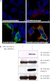
Arrestin and NHERF1 interact in living cells. A, detection of interaction between Myc-NHERF1 and GFP-arrestin-2 in HEK293 cells using DuoLink. Cells were transiently transfected with GFP-arrestin-2 and/or Myc-tagged NHERF1. Fixed and permeabilized cells were incubated with anti-GFP and anti-Myc primary antibodies. Secondary antibodies attached to PLA probes were incubated with the cells, prior to ligation and amplification steps as outlined under “Experimental Procedures.” Only when both probes bind in close proximity (<40 nm) is a PLA signal detected (shown in red), indicative of protein interaction. B, cells were transiently transfected with GFP-arrestin-2 and Myc-tagged NHERF1. Arrestin was immunoprecipitated (IP) with an anti-GFP antibody. Samples were resolved by SDS-PAGE and immunoblotted (IB) with an anti-Myc antibody to probe for associated NHERF1. Membranes were stripped and re-probed for total GFP-arrestin immunoprecipitated across samples. Inputs were immunoblotted with anti-GFP and anti-Myc antibodies to show efficient expression of transfected proteins across samples. Blots are representative of three independent experiments. Data shown are representative of three independent experiments. The scale bar represents 10 μm.
FIGURE 7.
FIGURE 7.
Potential mechanism of P2Y12 receptor internalization. 1) NHERF1 associates with the P2Y12 receptor in the absence of agonist via an interaction with the PDZ ligand (ETPM). 2) Receptor activation promotes GRK-dependent phosphorylation of C-tail and arrestin recruitment resulting in desensitization. 3) Arrestin binding to P2Y12 promotes PDZ ligand-independent NHERF1 recruitment to the receptor likely through direct interaction between arrestin and NHERF1. 4) Arrestin and NHERF1 both coordinate P2Y12 receptor internalization through a distinct population of clathrin-coated pits.

Similar articles

Cited by

References

    1. Wolfe B. L., Trejo J. (2007) Clathrin-dependent mechanisms of G protein-coupled receptor endocytosis. Traffic 8, 462–470 - PubMed
    1. Hanyaloglu A. C., von Zastrow M. (2008) Regulation of GPCRs by endocytic membrane trafficking and its potential implications. Annu. Rev. Pharmacol. Toxicol. 48, 537–568 - PubMed
    1. Marchese A., Paing M. M., Temple B. R., Trejo J. (2008) G protein-coupled receptor sorting to endosomes and lysosomes. Annu. Rev. Pharmacol. Toxicol. 48, 601–629 - PMC - PubMed
    1. Romero G., von Zastrow M., Friedman P. A. (2011) Role of PDZ proteins in regulating trafficking, signaling, and function of GPCRs. Means, motif, and opportunity. Adv. Pharmacol. 62, 279–314 - PMC - PubMed
    1. Cao T. T., Deacon H. W., Reczek D., Bretscher A., von Zastrow M. (1999) A kinase-regulated PDZ-domain interaction controls endocytic sorting of the β2-adrenergic receptor. Nature 401, 286–290 - PubMed

Publication types

MeSH terms

LinkOut - more resources