. 2012 May 20;13(7):642-50.
doi: 10.1038/ni.2304.

Notch-RBP-J signaling regulates the transcription factor IRF8 to promote inflammatory macrophage polarization

Affiliations

Notch-RBP-J signaling regulates the transcription factor IRF8 to promote inflammatory macrophage polarization

Haixia Xu et al. Nat Immunol. .

Abstract

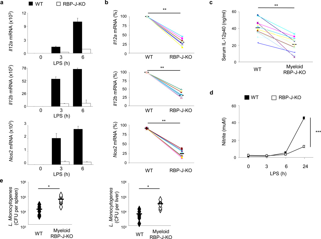
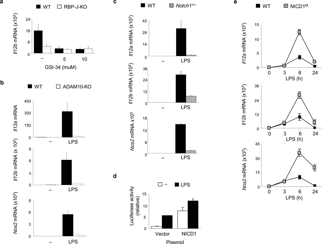
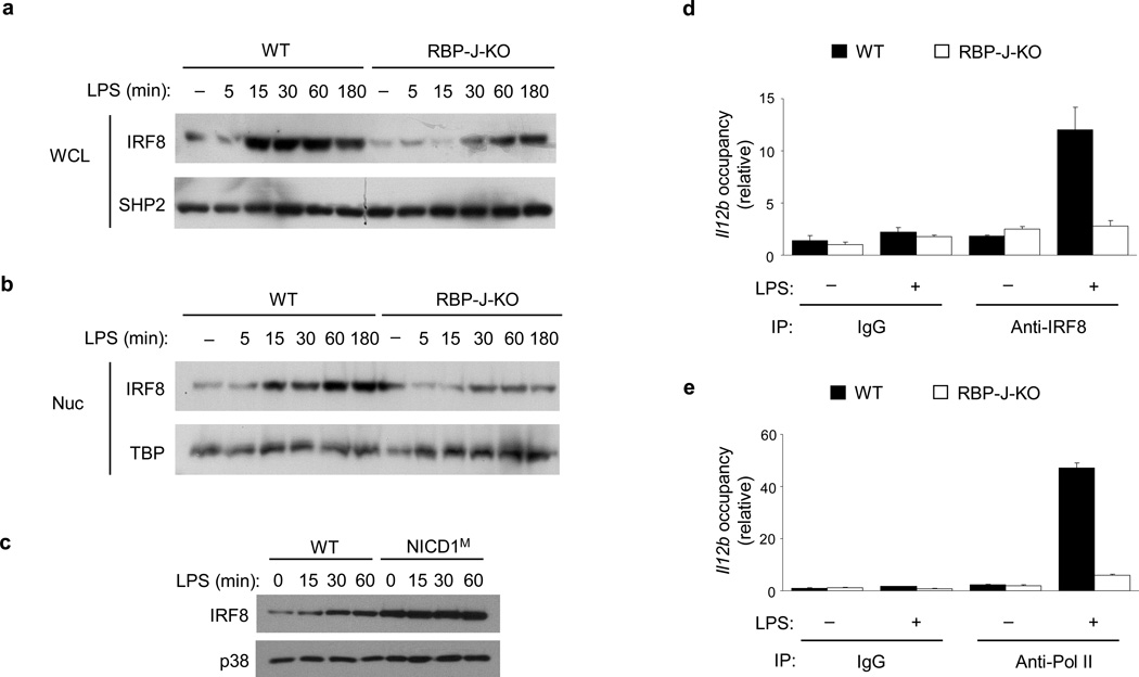
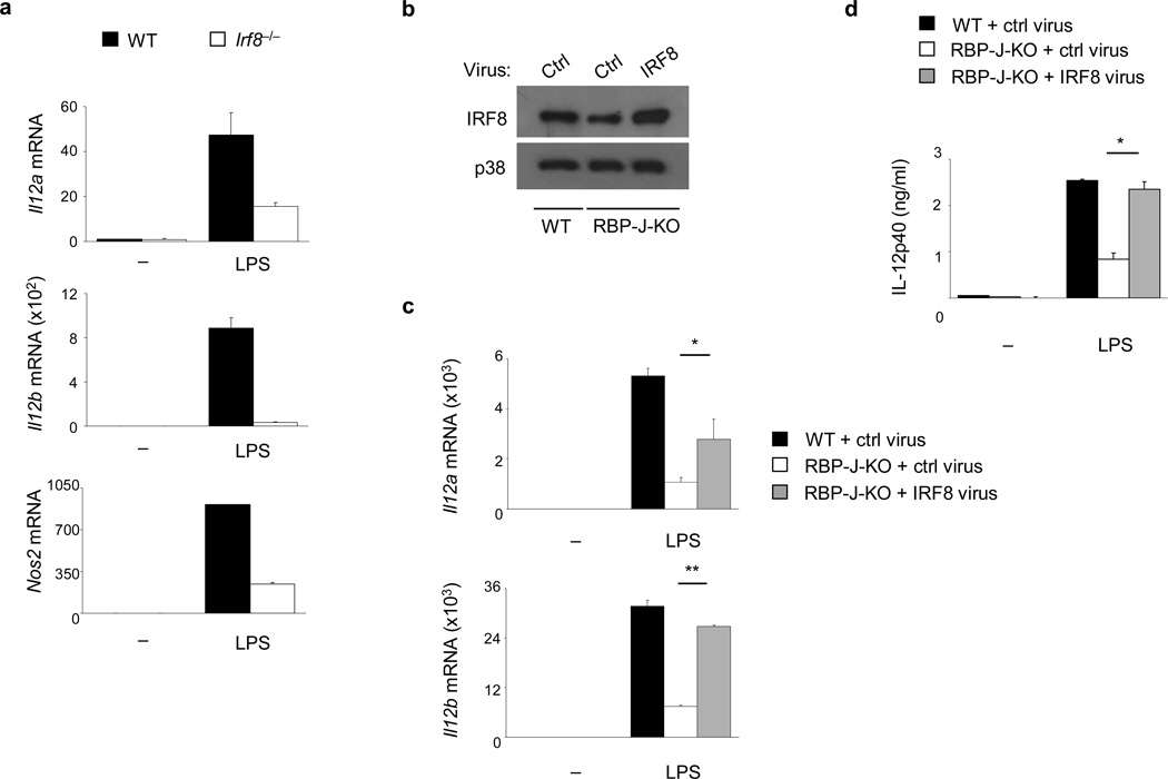
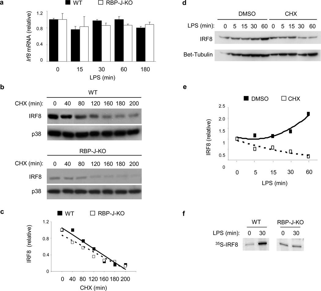
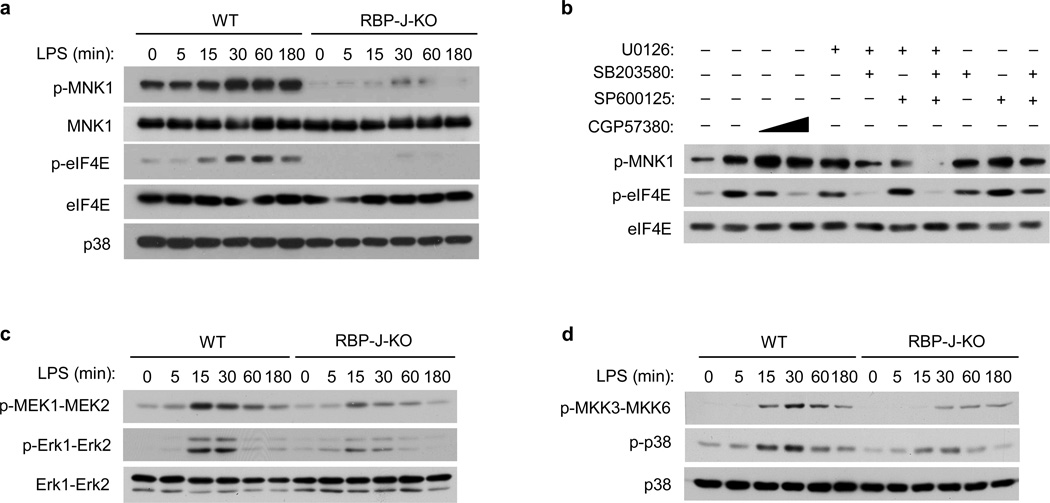
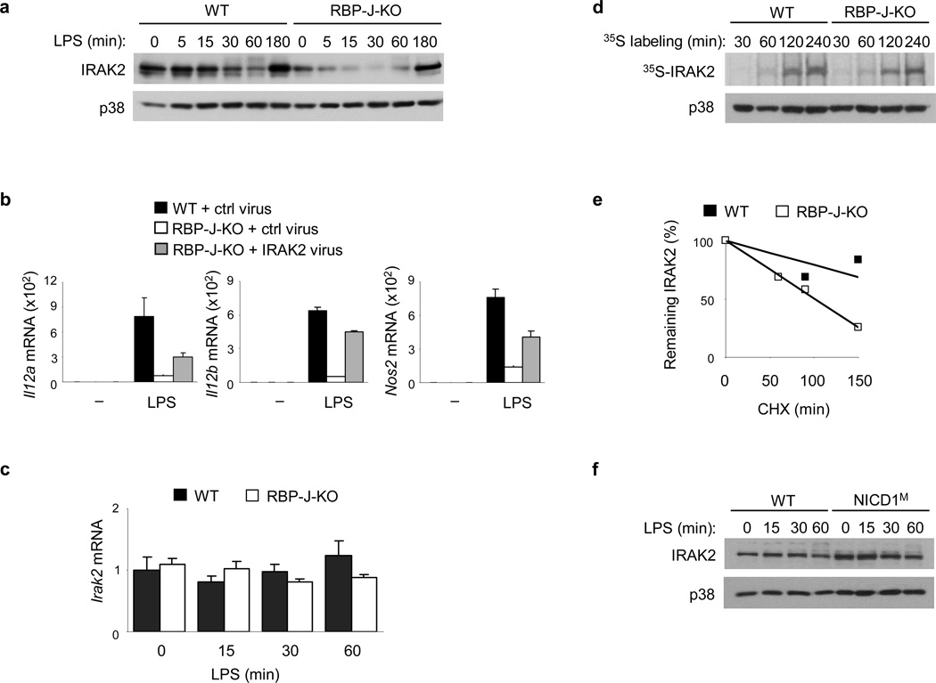
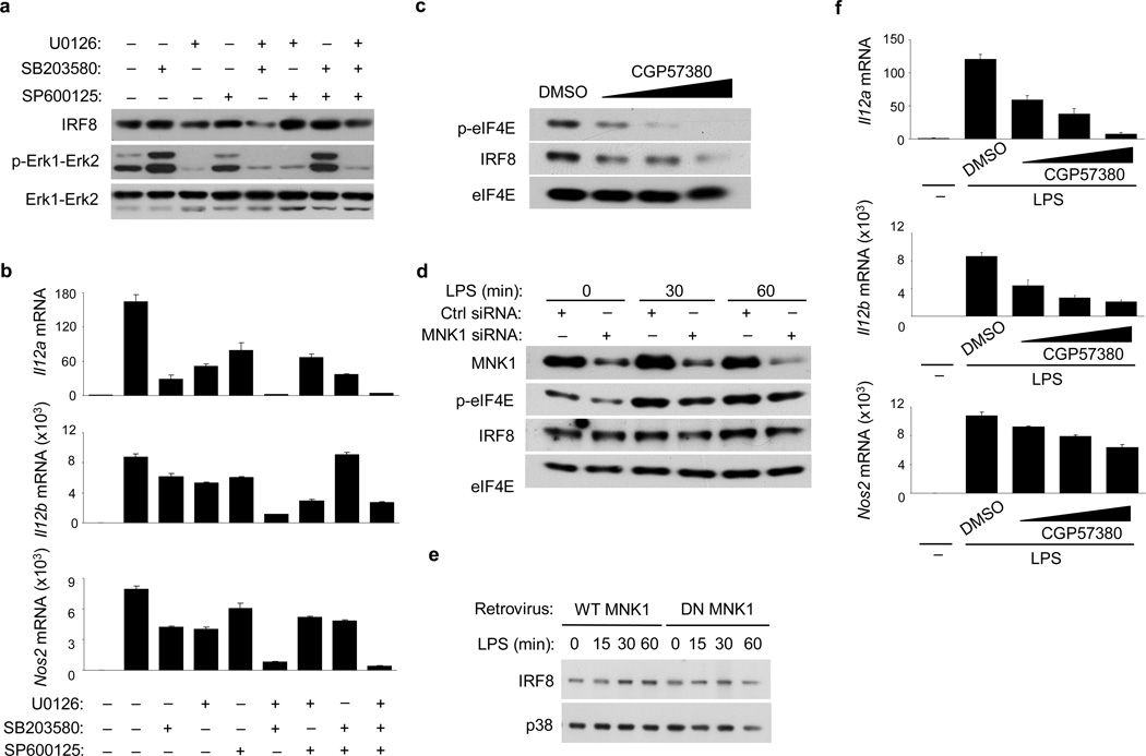
Emerging concepts suggest that the functional phenotype of macrophages is regulated by transcription factors that define alternative activation states. We found that RBP-J, the main nuclear transducer of signaling via Notch receptors, augmented Toll-like receptor 4 (TLR4)-induced expression of key mediators of classically activated M1 macrophages and thus of innate immune responses to Listeria monocytogenes. Notch-RBP-J signaling controlled expression of the transcription factor IRF8 that induced downstream M1 macrophage-associated genes. RBP-J promoted the synthesis of IRF8 protein by selectively augmenting kinase IRAK2-dependent signaling via TLR4 to the kinase MNK1 and downstream translation-initiation control through eIF4E. Our results define a signaling network in which signaling via Notch-RBP-J and TLRs is integrated at the level of synthesis of IRF8 protein and identify a mechanism by which heterologous signaling pathways can regulate the TLR-induced inflammatory polarization of macrophages.


Figures

Figure 1
Figure 1. RBP-J controls expression of prototypical M1 genes
(a) Quantitative PCR of mRNA expression of the indicated genes in BMDMs from Rbpjflox/flox, Mx1-Cre (RBP-J-KO) mice and Rbpj+/+, Mx1-Cre (wild-type; WT) littermate controls, stimulated with LPS (1 ng/ml) for the indicated periods. Unstimulated wild-type controls were set to 1. Results are normalized relative to Gapdh mRNA. Data are shown as means + SD of triplicate determinants. (b) Quantitative PCR of mRNA expression of the indicated genes in BMDMs from Rbpj+/+, Mx1-Cre (WT) and Rbpjflox/flox, Mx1-Cre (RBP-J-KO) mice, stimulated with LPS for 3 or 6 h. Data are represented as percentage of maximal mRNA expression in wild-type cells. Cumulative data from 6–12 independent experiments is shown. (c) ELISA of IL-12p40 expression in the serum of Rbpjflox/flox, LysM-Cre (RBP-J-KO) and Rbpj+/+, LysM-Cre (WT) mice, injected intraperitoneally with 200 µg of LPS, followed by blood collection 3 h later (n = 12 per group; average values are shown as horizontal bars). (d) NO release measured, as the NO metabolite nitrite, in the supernatants of Rbpj+/+, Mx1-Cre (WT) and Rbpjflox/flox, Mx1-Cre (RBP-J-KO) BMDMs, treated with LPS. Data shown are representative of three independent experiments. (e) Bacterial CFUs in the spleens and livers of Rbpjflox/flox, LysM-Cre (RBP-J-KO) or control Rbpj+/+, LysM-Cre (WT) bone marrow chimeras infected intravenously with 3 × 10³ L. monocytogenes strain 10403S and analyzed at day 3.5 post infection (n= 6 per group). * P<0.05, ** P<0.001, *** P<0.0001 (Student’s t test).
Figure 2
Figure 2. Induction of RBP-J-dependent M1 genes requires canonical Notch signaling
(a) Quantitative PCR of IL12b mRNA expression in BMDMs from Rbpj+/+, Mx1-Cre (wild-type; WT) and Rbpjflox/flox, Mx1-Cre (RBP-J-KO) paired littermates, pretreated with the γ-secretase inhibitor, GSI-34, for 48 h and then stimulated with LPS (1 ng/ml) for 3 h. (b) Quantitative PCR of mRNA expression of the indicated genes in BMDMs from wild-type (WT) or ADAM10-deficient (ADAM10-KO) BMDMs stimulated with LPS for 3 h. (c) Quantitative PCR of mRNA expression of the indicated genes in BMDMs from Notch1+/− mice and wild-type (WT) littermate controls, stimulated with LPS for 3 h. (d) Relative luciferase activity in lysates from RAW264.7 cells co-transfected with an Il12b reporter construct and a NICD1 expression plasmid or empty vector control, followed by stimulation for 6 h with LPS (1 µg/ml) at 36 h post-transfection. (e) Quantitative PCR of mRNA expression of the indicated genes in BMDMs from NICD1M mice and wild-type (WT) littermate controls, stimulated with LPS for the indicated periods. Data are shown as means + SD of triplicate determinants. Results shown are representative of at least three independent experiments.
Figure 3
Figure 3. RBP-J controls IRF8 expression and function
(a, b) Immunoblot analysis of IRF8 expression in whole cell lysates (WCL) (a) and nuclear (Nuc) extracts (b) of BMDMs from Rbpj+/+, Mx1-Cre (wild-type; WT) and Rbpjflox/flox, Mx1-Cre (RBP-J-KO) paired littermates, stimulated with LPS for the indicated periods. SHP2 (a) and TBP (b) served as loading controls. Data shown are representative of six independent experiments. (c) Immunoblot analysis of IRF8 expression in whole cell lysates of BMDMs from NICD1M mice and (wild-type) WT littermate controls, stimulated with LPS for the indicated periods. p38 served as a loading control. Data are representative of two independent experiments. (d, e) ChIP assays were performed with LPS-treated Rbpj+/+, Mx1-Cre (WT) and Rbpjflox/flox, Mx1-Cre (RBPJ-KO) macrophages, using antibodies against IRF8 (d) and Pol II (e). Occupancy was determined by qPCR amplification over the promoter region of Il12b. Data are shown as means + SD of triplicate determinants and are representative of three independent experiments.
Figure 4
Figure 4. IRF8 mediates RBP-J-dependent activation of M1 gene expression
(a) Quantitative PCR of mRNA expression of the indicated genes in BMDMs from WT and Irf8−/− mice, stimulated with LPS for 3 h. Data are representative of three independent experiments. (b) Immunoblot analysis of IRF8 expression in BMDMs from Rbpj+/+, Mx1-Cre (wild-type; WT) and Rbpjflox/flox, Mx1-Cre (RBP-J-KO) paired littermates transduced with an IRF8-expressing retrovirus or control (ctrl) virus. p38 served as a loading control. (c) Quantitative PCR assays of mRNA expression of the indicated genes in the virally transduced BMDMs from (b), stimulated for 6 h with LPS at 48 h post-transduction. (d) ELISA analysis of IL-12p40 expression in the transduced BMDMs from (b), stimulated for 6 h with LPS at 48 h post-transduction (d). Data are shown as means + SD of triplicate determinants and are representative of two independent experiments. *P<0.05, **P<0.001 (Student’s t test).
Figure 5
Figure 5. RBP-J promotes IRF8 protein synthesis
(a) Quantitative PCR of Irf8 mRNA in BMDMs from Rbpj+/+, Mx1-Cre (wild-type; WT) and Rbpjflox/flox, Mx1-Cre (RBP-J-KO) paired littermates, stimulated with LPS for the indicated periods. (b) Immunoblot analysis of IRF8 expression in whole cell lysates of BMDMs from Rbpj+/+, Mx1-Cre (WT) and Rbpjflox/flox, Mx1-Cre (RBP-J-KO) BMDMs, stimulated with LPS for 1 h and subsequently treated with the protein synthesis inhibitor, CHX (20 µg/ml) for the indicated periods. p38 served as a loading control. (c) Band intensities from (b) were quantitated using ImageJ software and normalized relative to the CHX 0 min conditions. (d) Immunoblot analysis of IRF8 expression in BMDMs from wild-type mice, pretreated with CHX (20 µg/ml) for 30 min and then activated with LPS for the indicated periods. β-tubulin served as a loading control. (e) Band intensities from (d) were quantitated using ImageJ software and normalized relative to the CHX 0 min conditions. (f) SDS-PAGE analysis of immunoprecipitated radiolabeled IRF8 in Rbpj+/+, Mx1-Cre (WT) and Rbpjflox/flox, Mx1-Cre (RBP-J-KO) BMDMs following metabolic labeling. Results shown are representative of at least three independent experiments.
Figure 6
Figure 6. RBP-J augments TLR4-induced activation of the MAPK-MNK1-eIF4E pathway
(a) Immunoblot analysis of the indicated proteins in whole cell lysates of BMDMs from Rbpj+/+, Mx1-Cre (wild-type; WT) and Rbpjflox/flox, Mx1-Cre (RBP-J-KO) paired littermates, stimulated with LPS for the indicated periods. p38 served as a loading control. (b) Immunoblot analysis of p-MNK1 and p-eIF4E in whole cell lysates from wild-type BMDMs, pretreated with DMSO vesicle control, a MNK inhibitor CGP57380, or MAPK inhibitors for 30 min and then stimulated with LPS for 60 min. Total eIF4E levels served as loading controls. (c & d) Immunoblot analysis of p-Erk1-Erk2 and p-MEK1-MEK2 (c) and p-p38 and p-MKK3-MKK6 (d) expression in BMDMs from Rbpj+/+, Mx1-Cre (WT) and Rbpjflox/flox, Mx1-Cre (RBP-J-KO) mice, stimulated with LPS for the indicated periods. Total Erk1-Erk2 (c) and total p38 (d) served as loading controls. Cumulative data from three independent experiments is shown.
Figure 7
Figure 7. Notch-RBP-J signaling regulates IRAK2 protein expression
(a) Immunoblot analysis of IRAK2 expression in BMDMs from Rbpj+/+, Mx1-Cre (wild-type; WT) and Rbpjflox/flox, Mx1-Cre (RBP-J-KO) mice, stimulated with LPS for the indicated periods. p38 served as a loading control. (b) Quantitative PCR of mRNA expression of the indicated genes in BMDMs from Rbpj+/+, Mx1-Cre (WT) and Rbpjflox/flox, Mx1-Cre (RBP-J-KO) mice, transduced with an IRAK2-expressing retrovirus, or a control (ctrl) virus, and stimulated for 6 h with LPS at 48 h post-transduction. (c) Quantitative PCR of Irak2 mRNA in BMDMs from Rbpj+/+, Mx1-Cre (WT) and Rbpjflox/flox, Mx1-Cre (RBP-J-KO) mice, stimulated with LPS for theindicated periods. (d) SDS-PAGE analysis of immunoprecipitated radiolabelled IRAK2 in BMDMs from Rbpj+/+, Mx1-Cre (WT) and Rbpjflox/flox, Mx1-Cre (RBP-J-KO) mice, following metabolic labeling for the indicated periods. Immunoblotted p38 in the whole cell lysates served as a loading control. (e) Quantitiation using ImageJ software, of IRAK2 expression in whole cell lysates of BMDMs from Rbpj+/+, Mx1-Cre (WT) and Rbpjflox/flox, Mx1-Cre (RBP-J-KO) mice, stimulated for 30 min with LPS followed by treatment with the protein synthesis inhibitor, CHX (20 µg/ml) for the indicated periods (f) Immunoblot analysis of IRAK2 expression in whole cell lyastes of BMDMs from NICD1M mice and wild-type (WT) littermate controls, stimulated with LPS for the indicated periods. Data are shown as means + SD of triplicate determinants (b, c). Data are representative of at least three independent experiments.
Figure 8
Figure 8. The MAPK-MNK1-eIF4E axis promotes IRF8 synthesis and M1 gene expression
(a) Immunoblot analysis of IRF8 and p-Erk1-Erk2 expression in BMDMs from wild-type mice, pretreated with DMSO vesicle control or MAPK inhibitors for 30 min and then stimulated with LPS for 60 min. Total Erk1-Erk2 levels served as loading controls. (b) Quantitative PCR of mRNA expression of the indicated genes in BMDMs from wild-type mice, pretreated with MAPK inhibitors and stimulated with LPS for 3 h. (c) Immunoblot analysis of p-eIF4E and IRF8 expression in wild-type BMDMs, pretreated with increasing doses of the MNK inhibitor CGP57380 or DMSO vesicle control for 30 min and then stimulated with LPS for 60 min. Total eIF4E levels served as loading controls. (d) Immunoblot for the indicated proteins in wild-type BMDMs, transfected with control (ctrl) non-targeting or MNK1-specific siRNA, and treated with LPS at 2 d post-transfection. (e) Immunoblot analysis of IRF8 expression in wild-type BMDMs, transduced with a retrovirus encoding wild-type (WT) MNK1 or a dominant negative (DN) MNK1 mutant and stimulated with LPS for the indicated periods following selection in puromycin-containing medium for 4 d. (f) Quantitative PCR of mRNA expression of the indicated genes in wild-type BMDMs, pretreated with increasing doses of CGP57380 or DMSO vesicle control for 30 min and then stimulated with LPS for 3 h. Data are representative of at least three independent experiments.

Similar articles

Cited by

References

    1. Mosser DM, Edwards JP. Exploring the full spectrum of macrophage activation. Nat. Rev. Immunol. 2008;8:958–969. - PMC - PubMed
    1. Krausgruber T, et al. IRF5 promotes inflammatory macrophage polarization and TH1-TH17 responses. Nat. Immunol. 2011;12:231–238. - PubMed
    1. Satoh T, et al. The Jmjd3-Irf4 axis regulates M2 macrophage polarization and host responses against helminth infection. Nat. Immunol. 2010;11:936–944. - PubMed
    1. Tailor P, et al. The feedback phase of type I interferon induction in dendritic cells requires interferon regulatory factor 8. Immunity. 2007;27:228–239. - PMC - PubMed
    1. Holtschke T, et al. Immunodeficiency and chronic myelogenous leukemia-like syndrome in mice with a targeted mutation of the ICSBP gene. Cell. 1996;87:307–317. - PubMed

Publication types

MeSH terms

Substances