

Comparison of several attachment methods for human iPS, embryonic and adipose-derived stem cells for tissue engineering
- PMID: 22610948
- PMCID: PMC4086291
- DOI: 10.1002/term.1499
Comparison of several attachment methods for human iPS, embryonic and adipose-derived stem cells for tissue engineering
Abstract
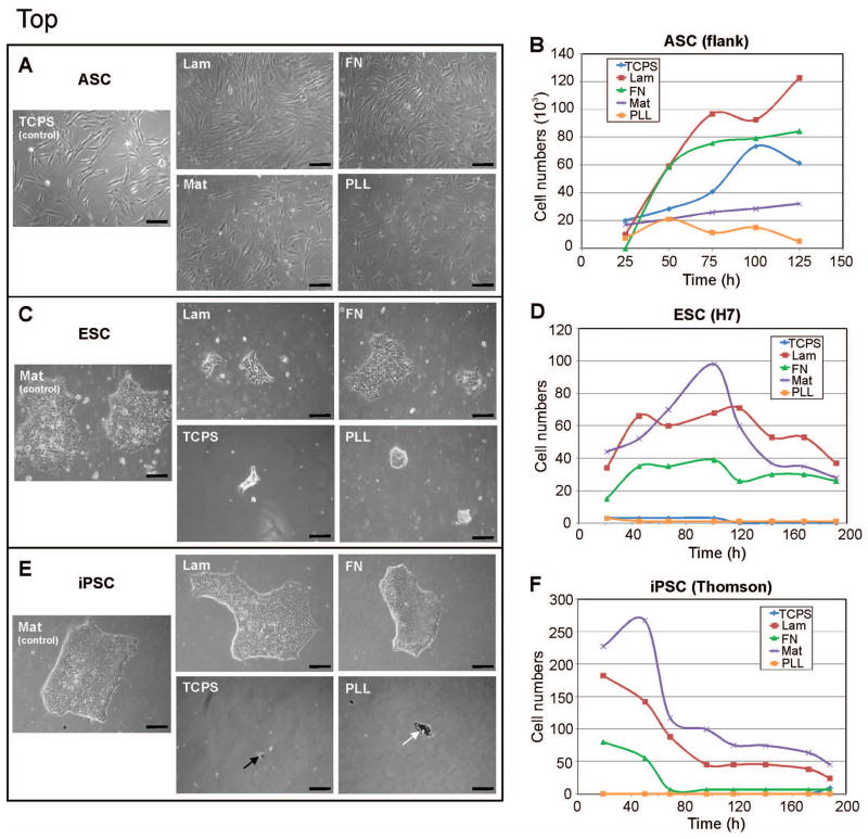
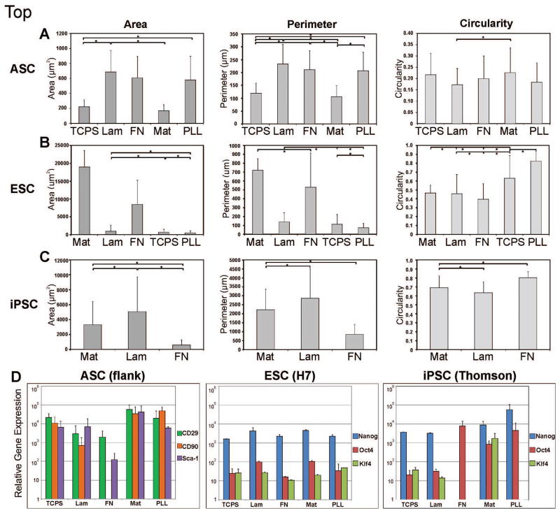
As actual stem cell application quickly approaches tissue engineering and regenerative medicine, aspects such as cell attachment to scaffolds and biomaterials become important and are often overlooked. Here, we compare the effects of several attachment proteins on the adhesion, proliferation and stem cell identity of three promising human stem cell types: human adipose-derived stem cells (hASCs), human embryonic stem cells (hESCs) and human induced pluripotent stem cells (hiPSCs). Traditional tissue culture polystyrene plates (TCPS), Matrigel (Mat), laminin (Lam), fibronectin (FN) and poly-L-lysine (PLL) were investigated as attachment protein surfaces. For hASCs typically cultured on TCPS, laminin resulted in the greatest cell attachment and proliferation with largest cell areas, indicating favourability by cell spreading. However, mesenchymal stem cell markers indicative of hASCs were slightly more expressed on surfaces with lowest cell attachment, corresponding to increased cell roundness, a newly observed attribute in hASCs possibly indicating a more stem cell-like character. hESCs preferred Matrigel as a feeder-free culture surface. Interestingly, hiPSCs favoured laminin over Matrigel for colony expansion, shown by larger cell colony area and perimeter lengths, although cell numbers and stem cell marker expression level remained highest on Matrigel. These data provide a practical reference guide for selecting a suitable attachment method for using human induced pluripotent, embryonic or adipose stem cells in tissue engineering and regenerative medicine applications.
Copyright © 2012 John Wiley & Sons, Ltd.
Conflict of interest statement
No competing financial interests exist.
Figures
References
-
- Braam SR, Zeinstra L, Litjens S, Ward-van Oostwaard D, van den Brink S, van Laake L, Lebrin F, Kats P, Hochstenbach R, Passier R, Sonnenberg A, Mummery CL. Recombinant vitronectin is a functionally defined substrate that supports human embryonic stem cell self-renewal via alphavbeta5 integrin. Stem Cells. 2008;26(9):2257–2265. - PubMed
-
- Domogatskaya A, Rodin S, Boutaud A, Tryggvason K. Laminin-511 but not -332, -111, or -411 enables mouse embryonic stem cell self-renewal in vitro. Stem Cells. 2008;26(11):2800–2809. - PubMed
-
- Hynes RO. Integrins: versatility, modulation, and signaling in cell adhesion. Cell. 1992;69(1):11–25. - PubMed
-
- Jokinen J, Dadu E, Nykvist P, Käpylä J, White DJ, Ivaska J, Vehviläinen P, Reunanen H, Larjava H, Häkkinen L, Heino J. Integrin-mediated cell adhesion to type I collagen fibrils. J Biol Chem. 2004;279(30):31956–31963. - PubMed
Publication types
MeSH terms
Grants and funding
LinkOut - more resources
Full Text Sources
Other Literature Sources
Research Materials
Miscellaneous
