. 2012 Aug;26(8):3118-26.
doi: 10.1096/fj.12-208991. Epub 2012 May 18.

Insulin resistance improves metabolic and contractile efficiency in stressed rat heart

Affiliations

Insulin resistance improves metabolic and contractile efficiency in stressed rat heart

Romain Harmancey et al. FASEB J. 2012 Aug.

Abstract

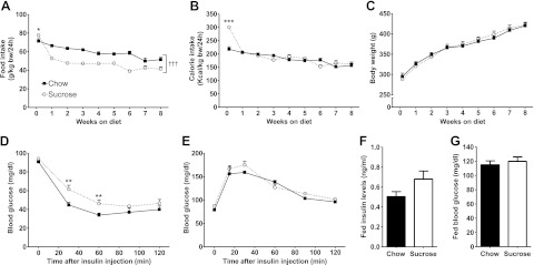
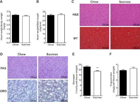
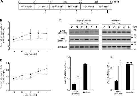
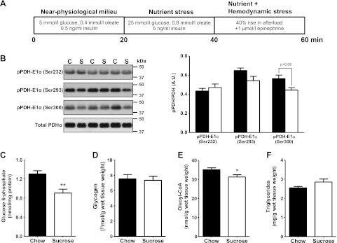
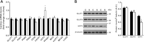
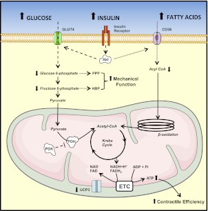
Insulin resistance is a prominent feature in heart failure, while hyperglycemia impairs cardiac contraction. We propose that decreased insulin-mediated glucose uptake by the heart preserves cardiac function in response to metabolic and hemodynamic stress. To test this hypothesis, we fed rats a high-sucrose diet (HSD). Energy substrate metabolism and cardiac work were determined ex vivo in a sequential protocol simulating metabolic and hemodynamic stress. Compared to chow-fed, control rats, HSD impaired myocardial insulin responsiveness and induced profound metabolic changes in the heart, characterized by reduced rates of glucose uptake (7.91 ± 0.30 vs. 10.73 ± 0.67 μmol/min/g dry weight; P<0.001) but increased rates of glucose oxidation (2.38 ± 0.17 vs. 1.50 ± 0.15 μmol/min/g dry weight; P<0.001) and oleate oxidation (2.29 ± 0.11 vs. 1.96 ± 0.12 μmol/min/g dry weight; P<0.05). Tight coupling of glucose uptake and oxidation and improved cardiac efficiency were associated with a reduction in glucose 6-phosphate and oleoyl-CoA levels, as well as a reduction in the content of uncoupling protein 3. Our results suggest that insulin resistance lessens fuel toxicity in the stressed heart. This calls for a new exploration of the mechanisms regulating substrate uptake and oxidation in the insulin-resistant heart.


Figures

Figure 1.
Figure 1.
The HSD-fed rat as a nonobese rodent model of insulin resistance. A) Food intake of chow-fed (solid squares with solid trace) and HSD-fed (open circles with dotted trace) rats was recorded over the first 24 h of feeding, and then weekly until the end of the protocol. *P < 0.05 and †††P < 0.001 vs. chow from 1 wk until 8 wk. B) Calorie intake was determined according to the energy content of the standard rodent chow (3.04 kcal/g) and of the HSD (3.90 kcal/g). ***P < 0.001 vs. chow at d 1. C) Body weight was measured for the same time points. Data are means ± se from 12 animals/group. D) An insulin tolerance test was performed starting 5 wk after the beginning of the feeding protocol by performing a subscapular subcutaneous injection of 0.5 U insulin/kg body weight in rats that were unfed for 18 h. Data are means ± se from 11 chow- and 9 HSD-fed rats. **P < 0.01 vs. chow. E) A glucose tolerance test was performed on rats that were unfed for 18 h after 5 wk on diet by administering 1g glucose/kg body weight by oral gavage of a 50% (w/v) glucose solution. Data are means ± se from 4 chow-fed and 5 HSD-fed rats. F, G) Fed plasma insulin levels (F) and fed blood glucose levels (G) were measured at time of sacrifice prior to heart perfusion. Data represent means ± se from 6 animals/group.
Figure 2.
Figure 2.
Effects of HSD on tissue structure and substrate storage in the heart. A, B) Cardiac size of chow-fed (solid bars) and HSD-fed (open bars) rats was assessed by normalizing heart weight to body weight (A) or heart weight to tibial length (B). C) Cardiac cell size and extracellular fibrosis were analyzed on H&E- and Masson's trichrome (MT)-stained cardiac tissue paraffin sections, respectively. D) Intracardiac glycogen and triglyceride levels were compared by PAS and oil red O (ORO) staining, respectively. E, F) Intracardiac glycogen (E) and triglycerides (F) were further quantified by enzymatic methods. Data represent means ± se from 6 animals/group. **P < 0.01 vs. chow.
Figure 3.
Figure 3.
Impaired insulin responsiveness in the working heart of HSD-fed rats. A) Isolated hearts were perfused in the working mode for 48 min in Krebs-Henseleit buffer supplemented with 5 mM glucose and 0.8 mM oleate. After allowing for stabilization of baseline parameters, the hearts were perfused in presence of 10−11 M insulin. The insulin concentration was then increased 10-fold every 8 min and up to 10−7 M. Cardiac function and myocardial glucose utilization rates were monitored 5 min after each change in the insulin concentration (arrows). B, C) Rates of glucose uptake (B) and glucose oxidation (C) for the perfused hearts of chow-fed (solid squares with solid trace; n=5) or HSD-fed (open circles with dotted trace; n=7) rats were measured for each concentration of insulin tested. D) Immunoblot analysis of phospho-Akt in hearts from chow-fed (C; solid bars) and sucrose-fed (S; open bars) rats. Akt phosphorylation at Thr308 and Ser473 was assessed at baseline in nonperfused hearts and in hearts that were perfused ex vivo with insulin with the protocol described in A. Data are shown as means ± se. *P < 0.05 vs. chow.
Figure 4.
Figure 4.
Increased flux through PDH and reduced levels of intermediary metabolites in the stressed hearts of HSD-fed rats. A) Isolated hearts from chow-fed (C; solid bars; n=11) or sucrose-fed (S; open bars; n=9) rats were perfused successively in 3 different conditions for a total of 60 min. The hearts were perfused with 5 mM glucose, 0.4 mM oleate, and 0.5 ng/ml insulin (near-physiological milieu) during the first 20 min, before being perfused with a buffer containing 25 mM glucose, 0.8 mM oleate, and 5 ng/ml insulin (metabolic stress) for the next 20 min. A hemodynamic stress (afterload raised from 100 to 140 cmH2O; 1 μM epinephrine) was superimposed to the metabolic stress for the last 20 min of perfusion. Cardiac function and myocardial rates of glucose and fatty acid oxidation were monitored every 5 min and averaged for each perfusion condition. Data are reported in Table 2. B) At the end of the perfusion protocol, the levels of inhibitory phosphorylation of PDH on serine residues 232, 293, and 300 were determined by immunoblot. C–F) Intracardiac levels of the glycolytic intermediate glucose 6-phosphate (C), glycogen (D), the fatty acid intermediate oleoyl-CoA (E), and triglycerides (F) were quantified from freeze-clamped, perfused heart tissue. All data are presented as means ± se. *P < 0.05, **P < 0.01 vs. chow.
Figure 5.
Figure 5.
Post-transcriptional down-regulation of UCP3 in the heart of HSD-fed rats. A) Total RNA was prepared from the hearts of chow-fed (solid bars) or HSD-fed (open bars) rats and analyzed by real-time PCR to determine the abundance of transcripts encoding metabolic enzymes. The proteins analyzed included the glucose transporters GLUT1 and GLUT4; the PDK isoforms 1, 2, and 4; the fatty acid transporters CD36 and fatty acid transporter 1 (FATP1); the uncoupling protein UCP3; peroxisome proliferator-activated receptor α (PPARα); malonyl-CoA decarboxylase (MCD); muscle carnitine palmitoyl transferase-1 (mCPT1); and the medium-chain and long-chain acyl-CoA dehydrogenases (MCAD and LCAD, respectively). B) Protein levels of GLUT1, GLUT4, and UCP3 were measured by immunoblot and normalized to β-tubulin levels. Data represent means ± se from 6 animals/group. *P < 0.05 vs. chow.
Figure 6.
Figure 6.
Proposed mechanisms leading to increased contractile performance for the insulin-resistant, stressed hearts of HSD-fed rats. In presence of hyperglycemia and hyperinsulinemia, impaired insulin-stimulated phosphorylation of Akt limits glucose transport into cardiac myocytes. Because insulin is known to stimulate the translocation of the fatty acid transporter CD36 at the sarcolemma (20), insulin resistance is also likely to limit fatty acid uptake in the heart of HSD-fed rats. Both rates of glucose and fatty acid oxidation are increased in response to an acute increase in workload. However, glucose oxidation increases more than fatty acid oxidation, and combines to an improved mitochondrial energy coupling (reduced UCP3 levels) to increase contractile efficiency. The energetic response of the stressed heart may have been preserved by the impairment of insulin-stimulated fatty acid uptake, and the reduction of fatty acid-mediated inhibition of glucose oxidation. The increased coupling between glucose uptake and glucose oxidation limits the accumulation of glycolytic intermediates and their rerouting into the pentose phosphate pathway (PPP) and the hexosamine biosynthetic pathway (HBP). This could increase the mechanical function of the heart by preserving the activity of contractile proteins and calcium homeostasis in cardiac myocytes. Dashed lines represent pathways with a reduced activity. Dotted lines represent possible consequences of insulin resistance that have not been tested directly in the present model.

References

    1. Ingelsson E., Sundstrom J., Arnlov J., Zethelius B., Lind L. (2005) Insulin resistance and risk of congestive heart failure. JAMA 294, 334–341 - PubMed
    1. Garvey W. T., Hardin D., Juhaszova M., Dominguez J. H. (1993) Effects of diabetes on myocardial glucose transport system in rats: implications for diabetic cardiomyopathy. Am. J. Physiol. 264, H837–H844 - PubMed
    1. Kannel W. B., Hjortland M., Castelli W. P. (1974) Role of diabetes in congestive heart failure: the Framingham study. Am. J. Cardiol. 34, 29–34 - PubMed
    1. Rubin J., Matsushita K., Ballantyne C. M., Hoogeveen R., Coresh J., Selvin E. (2012) Chronic hyperglycemia and subclinical myocardial injury. J. Am. Coll. Cardiol. 59, 484–489 - PMC - PubMed
    1. Rossetti L. (2004) Glucose toxicity: effect of chronic hyperglycemia on insulin action. In Diabetes Mellitus: A Fundamental and Clinical Text (LeRoith D., Taylor S. I., Olefsky J. M., eds) pp. 939–951, Lippincott Williams & Wilkins, Philadelphia

Publication types

MeSH terms

LinkOut - more resources