. 2012 Jun 15;188(12):6399-406.
doi: 10.4049/jimmunol.1102903. Epub 2012 May 18.

CXCL17 is a mucosal chemokine elevated in idiopathic pulmonary fibrosis that exhibits broad antimicrobial activity

Affiliations

CXCL17 is a mucosal chemokine elevated in idiopathic pulmonary fibrosis that exhibits broad antimicrobial activity

Amanda M Burkhardt et al. J Immunol. .

Abstract

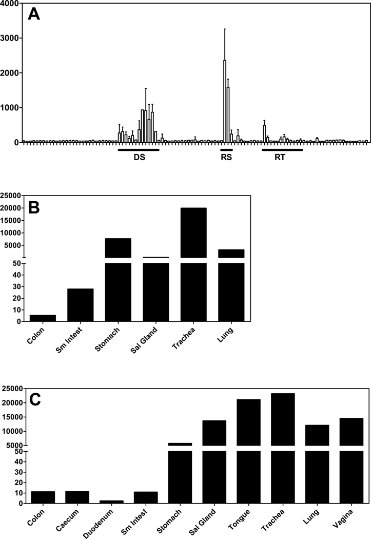
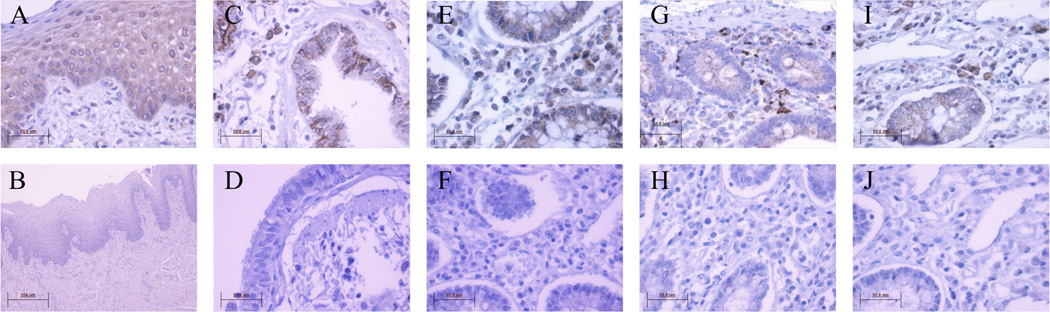
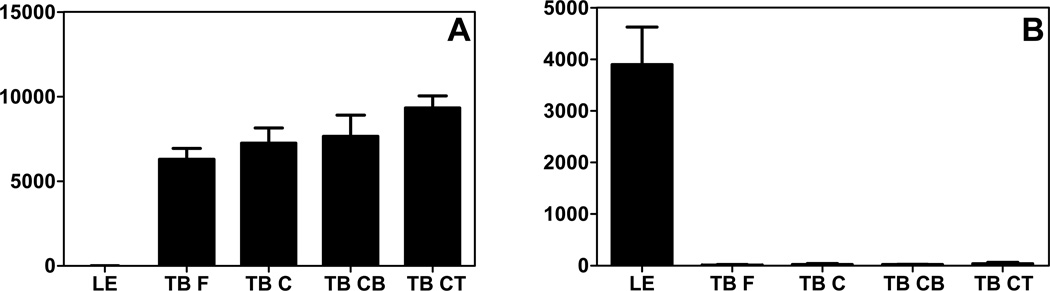
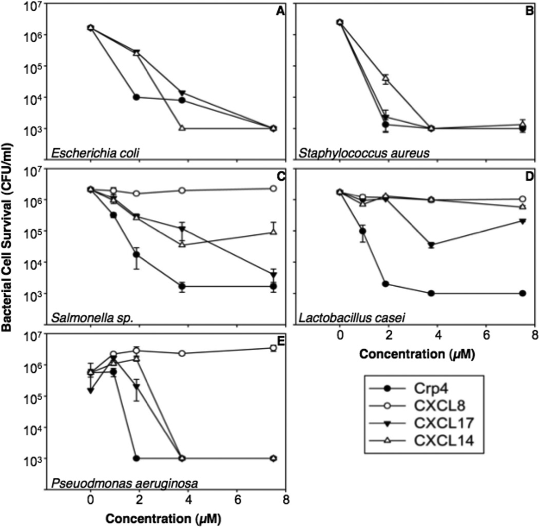
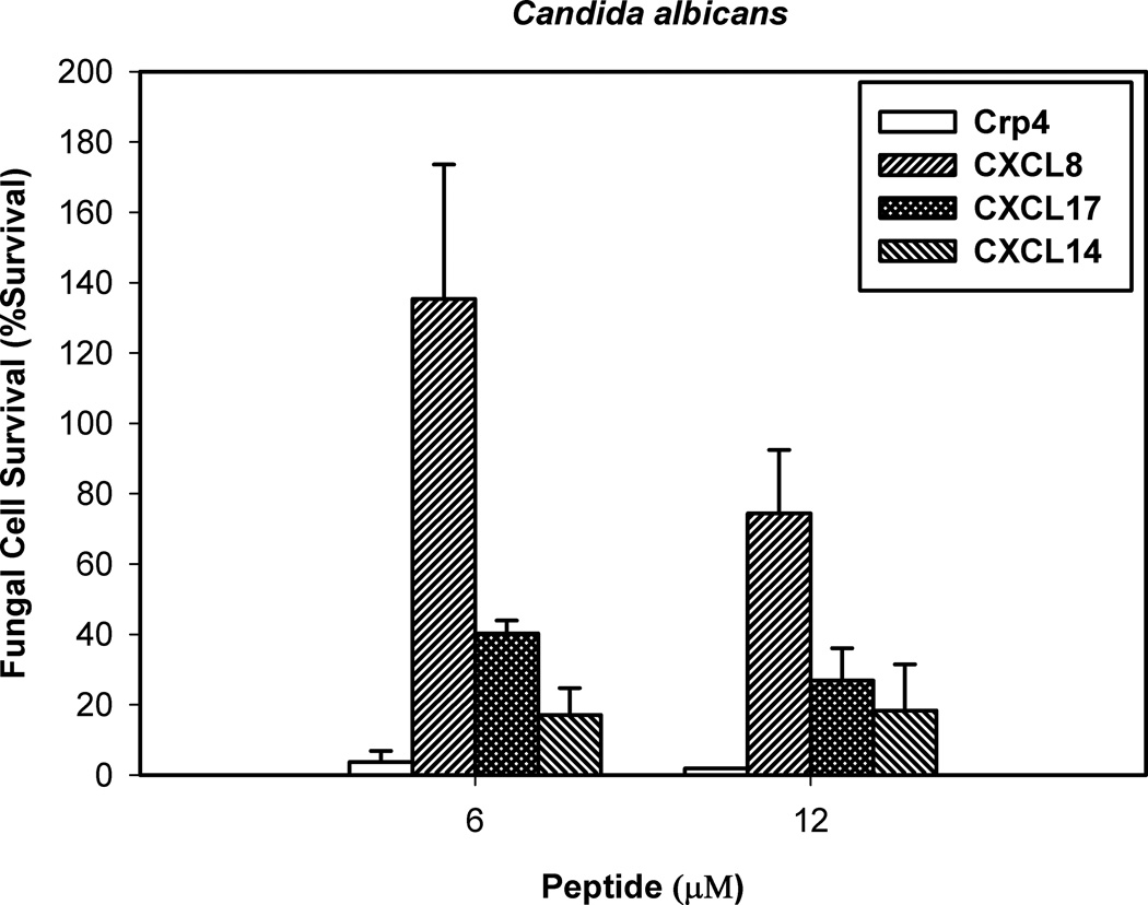
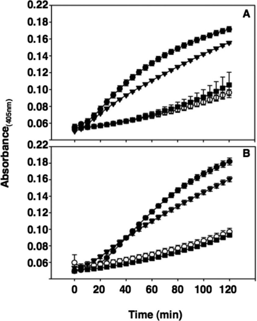
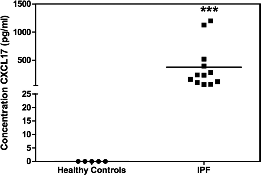
The mucosal immune network is a crucial barrier preventing pathogens from entering the body. The network of immune cells that mediates the defensive mechanisms in the mucosa is likely shaped by chemokines, which attract a wide range of immune cells to specific sites of the body. Chemokines have been divided into homeostatic or inflammatory depending upon their expression patterns. Additionally, several chemokines mediate direct killing of invading pathogens, as exemplified by CCL28, a mucosa-associated chemokine that exhibits antimicrobial activity against a range of pathogens. CXCL17 was the last chemokine ligand to be described and is the 17th member of the CXC chemokine family. Its expression pattern in 105 human tissues and cells indicates that CXCL17 is a homeostatic, mucosa-associated chemokine. Its strategic expression in mucosal tissues suggests that it is involved in innate immunity and/or sterility of the mucosa. To test the latter hypothesis, we tested CXCL17 for possible antibacterial activity against a panel of pathogenic and opportunistic bacteria. Our results indicate that CXCL17 has potent antimicrobial activities and that its mechanism of antimicrobial action involves peptide-mediated bacterial membrane disruption. Because CXCL17 is strongly expressed in bronchi, we measured it in bronchoalveolar lavage fluids and observed that it is strongly upregulated in idiopathic pulmonary fibrosis. We conclude that CXCL17 is an antimicrobial mucosal chemokine that may play a role in the pathogenesis of interstitial lung diseases.


Figures

Figure 1
Figure 1. Microarray data indicates that CXCL17 is a mucosal chemokine
(A) Mean expression values (y axis) from microarray data for 105 normal human tissues from the BIGE database (see Materials and Methods) (9, 11). Data are displayed across the x-axis grouped in organ systems. Y axis is the mean expression values. Highlighted organ systems, which have the highest expression of CXCL17, are: DS, digestive system; RS, respiratory system; RT, reproductive tract. The values for the tissues with the highest expression of CXCL17 are shown in Table 1. For a complete list of tissues included in the BIGE database consult (11). (B & C). Q-PCR in human and mouse mucosal tissue confirms that CXCL17 is highly expressed in mucosal tissues. Q-PCR with human (B) and mouse (C) CXCL17 specific primers was used to detect the expression of CXCL17 in each of the mucosal tissues (x axis). Expression Units of CXCL17 in mouse mucosal tissues (y axis) are based on Ct values averaged for multiple samples. Tissues were harvested from normal C57BL/6 mice. Expression for each tissue represents averaged expression for tissues collected from two different animals. Sm Intest, small intestine; Sal Gland, salivary gland.
Figure 2
Figure 2. CXCL17 exhibits specific expression patterns in several human mucosal tissues
Positive staining for CXCL17 was detected in epithelial cells of the lingual tongue mucosa (A), the luminal facing respiratory mucosa (C), and the lamina propria (LP) colon mucosa (E). The respective isotype controls are shown in (B), (D), and (F). Additionally, discrete cells within each of these mucosal sites stained positive for CXCL17 (A, C, E). Staining with cell type specific markers, in addition to morphological analysis (supplementary figure 1), confirmed that these are CD68+ macrophages (G) and CD138+ plasma cells (I) within each of the mucosal sites analyzed (G and I are in the colon LP, other macrophage and plasma cell staining in tongue and respiratory mucosa are data not shown). The respective isotype controls are shown in (H) and (J). All images was captured at 40X magnification expect Figure B which was captured at 10X.
Figure 3
Figure 3. CXCL14 and CXCL17 show mutually exclusive expression in the tongue
CXCL14 and CXCL17 microarray gene expression data from either lingual epithelium or taste buds. This analysis was performed using a tongue gene expression database described previously (10). (A) CXCL14 is only expressed in the samples derived from taste buds (TB F, fungiform; TB C, circumvallate; TB CB, circumvallate base; TB CT, circumvallate top). (B) Conversely, CXCL17 is only expressed in lingual epithelium (LE). X axis: discrete tongue tissue; Y axis: mean expression values. All samples were obtained from a variable number of healthy donors depending on the tissue: lingual epithelium, 10 donors; fungiform taste buds, 6 donors; circumvallate, 4 donors; circumvallate base, 4 donors; circumvallate top, 4 donors. The data is shown as averages of data from all donors and error bars denote standard deviation of the mean.
Figure 4
Figure 4. CXCL17 has broad spectrum bacteriocidal activity
Exponentially growing Escherichia coli (A), Staphylococcus aureus (B), Salmonella enterica serovar Typhimurium 14028s (C), Lactobacillus casei (D), Pseudomonas aeruginosa (E), were exposed to chemokines at 37°C in 50 µl of PIPES-TSB buffer for 1 h (see Materials and Methods). Following peptide exposure, the bacteria were spread on TSB-agar plates and incubated overnight at 37°C. Surviving bacteria were counted as CFU/ml at each concentration and values at or below 1 × 10³ CFU/ml mean that no detectable colonies were observed. Except for E. coli, assays were performed in triplicate and error bars denote standard deviation of the mean.
Figure 5
Figure 5. CXCL17 is active against the opportunistic fungal pathogen C. albicans
Log-phase C. albicans were exposed to chemokines at 6 and 12 µM protein concentrations at 37°C in 50 µl of PIPES-SAB buffer for 1 h. Following peptide exposure, the bacteria were spread on SAB-agar plates and incubated overnight at 37°C. Surviving Candida colonies were counted as CFU/ml at each concentration and values at or below 1 × 10³ CFU/ml mean no detectable colonies were observed. Assays were performed in triplicate and error bars denote standard deviation of the mean.
Figure 6
Figure 6. CXCL17 induces permeabilization of live E. coli membranes as measured by ONPG conversion
Log-phase E. coli ML35 cells were exposed to either 3.75 µM (A) or 1.625 µM (B) Crp4, CXCL17, CXCL8, and ProCryptdin4 in the presence of 2-nitrophenyl β-D-galactopyranoside (ONPG) for 2 h at 37°C. β-galactosidase mediated hydrolysis of ONPG was measured at A405. Symbols: Crp4, -●-; CXCL8, -○-; CXCL17, - ▼-; and, ProCrp4, -■-.
Figure 7
Figure 7. CXCL17 is elevated in BALf samples of patients diagnosed with IPF compared to healthy controls
CXCL17 was detected in human BALf samples using a CXCL17-specific sandwich ELISA. The BALf samples were concentrated to 10X prior to ELISA analysis. Recombinant human CXCL17 was used as the standard. CXCL17 was below the level of detection in healthy samples, but was detected at robust levels in samples from patients previously diagnosed with IPF. The CXCL17 levels in the IPF samples were statistically significant (indicated by ***) compared to healthy samples (p=0.01). Assays were performed in duplicate. X axis: samples; Y axis: concentration of CXCL17 (pg/ml).

References

    1. Zlotnik A, Yoshie O, Nomiyama H. The chemokine and chemokine receptor superfamilies and their molecular evolution. Genome Biol. 2006;7:243. - PMC - PubMed
    1. Van Der Meer P, Goldberg SH, Fung KM, Sharer LR, Gonzalez-Scarano F, Lavi E. Expression pattern of CXCR3, CXCR4, and CCR3 chemokine receptors in the developing human brain. J Neuropathol Exp Neurol. 2001;60:25–32. - PubMed
    1. Li M, Yu J, Li Y, Li D, Yan D, Ruan Q. CXCR4+ progenitors derived from bone mesenchymal stem cells differentiate into endothelial cells capable of vascular repair after arterial injury. Cell Reprogram. 2010;12:405–415. - PubMed
    1. Kunkel EJ, Campbell JJ, Haraldsen G, Pan J, Boisvert J, Roberts AI, Ebert EC, Vierra MA, Goodman SB, Genovese MC, Wardlaw AJ, Greenberg HB, Parker CM, Butcher EC, Andrew DP, Agace WW. Lymphocyte CC chemokine receptor 9 and epithelial thymus-expressed chemokine (TECK) expression distinguish the small intestinal immune compartment: Epithelial expression of tissue-specific chemokines as an organizing principle in regional immunity. J Exp Med. 2000;192:761–768. - PMC - PubMed
    1. Onai N, Kitabatake M, Zhang YY, Ishikawa H, Ishikawa S, Matsushima K. Pivotal role of CCL25 (TECK)-CCR9 in the formation of gut cryptopatches and consequent appearance of intestinal intraepithelial T lymphocytes. Int Immunol. 2002;14:687–694. - PubMed

Publication types

MeSH terms