. 2012 Jun 15;188(12):6145-55.
doi: 10.4049/jimmunol.1103660. Epub 2012 May 18.

Development of mature and functional human myeloid subsets in hematopoietic stem cell-engrafted NOD/SCID/IL2rγKO mice

Affiliations

Development of mature and functional human myeloid subsets in hematopoietic stem cell-engrafted NOD/SCID/IL2rγKO mice

Satoshi Tanaka et al. J Immunol. .

Abstract

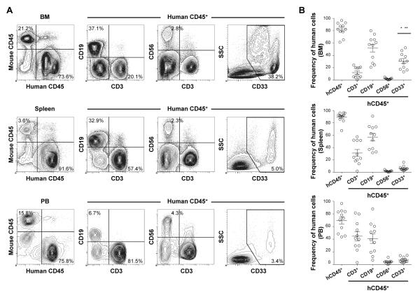
Although physiological development of human lymphoid subsets has become well documented in humanized mice, in vivo development of human myeloid subsets in a xenotransplantation setting has remained unevaluated. Therefore, we investigated in vivo differentiation and function of human myeloid subsets in NOD/SCID/IL2rγ(null) (NSG) mouse recipients transplanted with purified lineage(-)CD34(+)CD38(-) cord blood hematopoietic stem cells. At 4-6 mo posttransplantation, we identified the development of human neutrophils, basophils, mast cells, monocytes, and conventional and plasmacytoid dendritic cells in the recipient hematopoietic organs. The tissue distribution and morphology of these human myeloid cells were similar to those identified in humans. After cytokine stimulation in vitro, phosphorylation of STAT molecules was observed in neutrophils and monocytes. In vivo administration of human G-CSF resulted in the recruitment of human myeloid cells into the recipient circulation. Flow cytometry and confocal imaging demonstrated that human bone marrow monocytes and alveolar macrophages in the recipients displayed intact phagocytic function. Human bone marrow-derived monocytes/macrophages were further confirmed to exhibit phagocytosis and killing of Salmonella typhimurium upon IFN-γ stimulation. These findings demonstrate the development of mature and functionally intact human myeloid subsets in vivo in the NSG recipients. In vivo human myelopoiesis established in the NSG humanized mouse system may facilitate the investigation of human myeloid cell biology including in vivo analyses of infectious diseases and therapeutic interventions.


Figures

FIGURE 1
FIGURE 1
Development of human acquired and innate immunity in NSG recipients following transplantation of human CB HSCs. A, Representative sets of flow cytometry contour plots demonstrating the development of human CD45+ hematopoietic cells, hCD3+ T cells, hCD19+ B cells, hCD56+ NK cells and hCD33+ myeloid cells in the BM, spleen and PB of an NSG recipient. B, Human CD45+ hematopoietic chimerism and the frequencies of hCD3+ T, hCD19+ B, hCD33+ myeloid cells (n = 11 each, frequency of myeloid cells in BM compared with spleen; *p < 0.0001 and PB; **p < 0.0003) and hCD56+ NK (n = 9 each) cells in the BM, spleen and PB of NSG recipients at 4 to 6 months post-transplantation are summarized.
FIGURE 2
FIGURE 2
Development of human myeloid lineages in NSG recipients. A, Representative flow cytometry contour plots demonstrating differentiation of human HLA-DR granulocytes and HLA-DR+ APCs in the BM and spleen of an NSG recipient. B, The frequencies of human neutrophils (Neu), monocytes (Mo), cDCs, mast cells (Mast), basophils (Baso) and pDCs in the BM and spleen of NSG recipients are summarized (n = 10). C, In the humanized mouse BM and spleen, two distinct subsets of DCs, BDCA-1+ DCs and BDCA-3+ DCs were identified in HLA-DR+CD33+CD11c+ conventional DCs. Frequencies of the two DC subsets within BM and spleen hCD45+CD33+ cells are shown (BM; n=9, *p = 0.007, significant differences between cDCs, spleen; n=6, **p = 0.046). D, Human myeloid cells isolated by cell sorting of recipient BM demonstrate characteristic morphological features on May-Grünwald-Giemsa stain.
FIGURE 3
FIGURE 3
Expression of cytokine receptors on human myeloid cells in NSG recipients. A-C, Representative flow cytometry contour plots demonstrating the expression of IFN-γR, G-CSFR, GM-CSFR, and M-CSFR by CB hCD45+CD33+ myeloid cells (upper) and by hCD45+CD33+ myeloid cells derived from humanized NSG BM (lower). Contour plots for isotype control Ig are also shown. D, Expression of each cytokine receptor within hCD45+CD33+ cells is summarized (CB; n = 5, humanized NSG BM; n = 5).
FIGURE 4
FIGURE 4
Human myeloid lineage cells developing in NSG recipients demonstrate cytokine responses in vitro and in vivo. A, B, Phosphorylation of STAT1, STAT3, STAT4, STAT5, and STAT6 in human neutrophils and monocytes derived from a NSG recipient BM following in vitro stimulation with rhGM-CSF (A) and with rhG-CSF (B) was measured by flow cytometry. C, D, Results from three independent experiments using three different recipients are summarized. E, Heatmap representation of STAT phosphorylation in human neutrophils and monocytes in an NSG recipient BM following in vitro cytokine treatment relative to PBS exposure is shown. F, Representative flow cytometry contour plots demonstrating expansion of myeloid lineage cells in the PB of an NSG recipient in response to in vivo recombinant human G-CSF administration. Frequencies of hCD45+CD15+CD33low and hCD45+CD15−/lowCD33+ myeloid cells were increased following in vivo rhG-CSF treatment in PB of NSG recipients for 5 days.
FIGURE 5
FIGURE 5
Expression of TLRs and response to TLR adjuvant by humanized mouse-derived myeloid subsets. TLR expression is analyzed in the granulocytes, monocytes and cDCs derived from the humanized NSG recipient BM. A, B, Expression of TLR2 and TLR4 in neutrophils, monocytes, BDCA-1+ DCs, and BDCA-3+ DCs was analyzed by flow cytometry. C, At different time points after the injection of 15 μg LPS into humanized NSG recipients, human specific cytokine levels in plasma were evaluated by cytometric bead array (n = 3).
FIGURE 6
FIGURE 6
Human monocytes/macrophages developing in NSG recipient lung demonstrate phagocytosis of micro-particles. A, Representative contour plots demonstrating the reconstitution of human myeloid cells in the lungs of an NSG recipient. Human CD45+ cells within lung cell populations were analyzed by CD33, HLA-DR, CD14, CD11c, BDCA-1/3, and CD15 to identify monocytes/macrophages ( Mo/Mϕ), cDCs, and neutrophils (Neu). B, The frequencies of human neutrophils (Neu), monocytes/macrophages (Mo/Mϕ), cDCs, mast cells (Mast) and basophils (Baso) within hCD45+CD33+ NSG recipient lung are summarized (n = 8). C, A set of representative flow cytometry plots demonstrating the presence of hCD45+CD33+fluorescent beads+ cells. D, Summary of the frequency of hCD45+CD33+ fluorescent bead+ cells in NSG recipient lung cell populations incubated at 37°C and at 4°C (control), respectively, with fluorescent beads (lung; n = 6, BM; n = 4, *p = 0.001, **p = 0.01). E, Confocal imaging of FACS-purified hCD45+CD33+fluorescent beads+ cells derived from NSG recipient lung cell populations show internalization of fluorescent beads (green) within hCD45 (purple)-expressing human myeloid cells.
FIGURE 7
FIGURE 7
Cytotoxicity against S. typhimurium by IFN-γ activated human monocytes/macrophages developing in NSG recipient. A, Within mononuclear cell gate, PI viable, hCD45+LinCD11b+ cells were purified from the BM of humanized NSG recipients. Purified BM monocytes/macrophages were stimulated with or without supplementation of 1000 U/mL human IFN-γ for 24h and then infected with S. typhimurium at 20 MOI. B, Intracellular CFU was counted at 3 and 12 hours post-infection (n = 5, *p = 0.023 compared with non-stimulated).

Similar articles

Cited by

References

    1. Mosier DE, Gulizia RJ, Baird SM, Wilson DB. Transfer of a functional human immune system to mice with severe combined immunodeficiency. Nature. 1988;335:256–259. - PubMed
    1. McCune JM, Namikawa R, Kaneshima H, Shultz LD, Lieberman M, Weissman IL. The SCID-hu mouse: murine model for the analysis of human hematolymphoid differentiation and function. Science. 1988;241:1632–1639. - PubMed
    1. Greiner DL, Hesselton RA, Shultz LD. SCID mouse models of human stem cell engraftment. Stem Cells. 1998;16:166–177. - PubMed
    1. Shultz LD, Schweitzer PA, Christianson SW, Gott B, Schweitzer IB, Tennent B, McKenna S, Mobraaten L, Rajan TV, Greiner DL, et al. Multiple defects in innate and adaptive immunologic function in NOD/LtSz-scid mice. J Immunol. 1995;154:180–191. - PubMed
    1. Takenaka K, Prasolava TK, Wang JC, Mortin-Toth SM, Khalouei S, Gan OI, Dick JE, Danska JS. Polymorphism in Sirpa modulates engraftment of human hematopoietic stem cells. Nature Immunology. 2007;8:1313–1323. - PubMed

Publication types

Substances