

Identification and characterization of the mammalian association and actin-nucleating domains in the Rickettsia conorii autotransporter protein, Sca2
- PMID: 22612237
- PMCID: PMC3422371
- DOI: 10.1111/j.1462-5822.2012.01815.x
Identification and characterization of the mammalian association and actin-nucleating domains in the Rickettsia conorii autotransporter protein, Sca2
Abstract
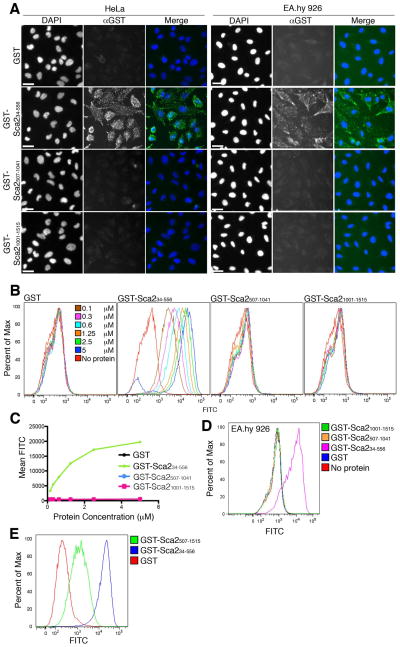
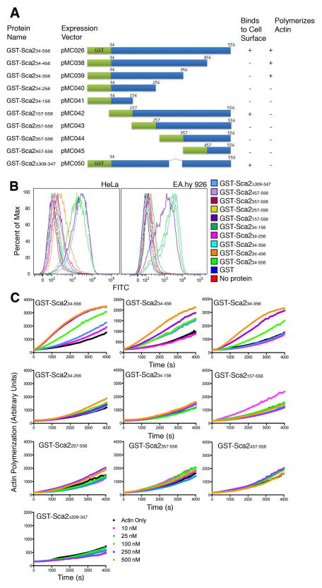
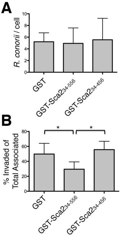
Establishment of infection by spotted fever group rickettsial species is dependent on the ability of these bacteria to adhere to and invade the host endothelium. Recent studies have attributed these processes to a handful of rickettsial surface proteins from the surface cell antigen (sca) family of autotransporters. A rickettsial autotransporter from Rickettsia conorii, Sca2, has been shown to be sufficient to mediate both adherence and invasion of human endothelial cells and to participate in intracellular actin-based motility. Here we identify a region of Sca2 capable of interacting with the mammalian cell surface and show that this function of Sca2 is independent and separable from its actin nucleation activity. Furthermore, pre-incubation of mammalian cells with the Sca2 mammalian association region prior to R. conorii infection can competitively inhibit rickettsial invasion, suggesting that Sca2 plays an important role in the initial interaction with mammalian cells. Together, our results demonstrate that the Sca2 autotransporter protein in R. conorii contains distinct functional domains that likely are involved in mediating cellular interactions at the plasma membrane and the host cytosol.
© 2012 Blackwell Publishing Ltd.
Figures
Similar articles
-
The Rickettsia conorii autotransporter protein Sca1 promotes adherence to nonphagocytic mammalian cells.Infect Immun. 2010 May;78(5):1895-904. doi: 10.1128/IAI.01165-09. Epub 2010 Feb 22. Infect Immun. 2010. PMID: 20176791 Free PMC article.
-
The Sca2 autotransporter protein from Rickettsia conorii is sufficient to mediate adherence to and invasion of cultured mammalian cells.Infect Immun. 2009 Dec;77(12):5272-80. doi: 10.1128/IAI.00201-09. Epub 2009 Oct 5. Infect Immun. 2009. PMID: 19805531 Free PMC article.
-
Disruption of the Rickettsia rickettsii Sca2 autotransporter inhibits actin-based motility.Infect Immun. 2010 May;78(5):2240-7. doi: 10.1128/IAI.00100-10. Epub 2010 Mar 1. Infect Immun. 2010. PMID: 20194597 Free PMC article.
-
Progress in rickettsial genome analysis from pioneering of Rickettsia prowazekii to the recent Rickettsia typhi.Ann N Y Acad Sci. 2005 Dec;1063:13-25. doi: 10.1196/annals.1355.003. Ann N Y Acad Sci. 2005. PMID: 16481486 Review.
-
[Emerging rickettsioses].Parassitologia. 2004 Jun;46(1-2):123-6. Parassitologia. 2004. PMID: 15305700 Review. Italian.
Cited by
-
A new role for host annexin A2 in establishing bacterial adhesion to vascular endothelial cells: lines of evidence from atomic force microscopy and an in vivo study.Lab Invest. 2019 Nov;99(11):1650-1660. doi: 10.1038/s41374-019-0284-z. Epub 2019 Jun 28. Lab Invest. 2019. PMID: 31253864 Free PMC article.
-
Which Way In? The RalF Arf-GEF Orchestrates Rickettsia Host Cell Invasion.PLoS Pathog. 2015 Aug 20;11(8):e1005115. doi: 10.1371/journal.ppat.1005115. eCollection 2015 Aug. PLoS Pathog. 2015. PMID: 26291822 Free PMC article.
-
Differences in Intracellular Fate of Two Spotted Fever Group Rickettsia in Macrophage-Like Cells.Front Cell Infect Microbiol. 2016 Jul 29;6:80. doi: 10.3389/fcimb.2016.00080. eCollection 2016. Front Cell Infect Microbiol. 2016. PMID: 27525249 Free PMC article.
-
Genomic Features of Rickettsia heilongjiangensis Revealed by Intraspecies Comparison and Detailed Comparison With Rickettsia japonica.Front Microbiol. 2019 Dec 6;10:2787. doi: 10.3389/fmicb.2019.02787. eCollection 2019. Front Microbiol. 2019. PMID: 31866968 Free PMC article.
-
Increased talin-vinculin spatial proximities in livers in response to spotted fever group rickettsial and Ebola virus infections.Lab Invest. 2020 Aug;100(8):1030-1041. doi: 10.1038/s41374-020-0420-9. Epub 2020 Apr 1. Lab Invest. 2020. PMID: 32238906 Free PMC article.
References
-
- Blanc G, Ngwamidiba M, Ogata H, Fournier PE, Claverie JM, Raoult D. Molecular evolution of rickettsia surface antigens: evidence of positive selection. Mol Biol Evol. 2005;22:2073–2083. - PubMed
Publication types
MeSH terms
Substances
Grants and funding
LinkOut - more resources
Full Text Sources
Research Materials
