

Pyonephrosis and urosepsis in a 41-year old patient with spina bifida: Case report of a preventable death
- PMID: 22613462
- PMCID: PMC3407709
- DOI: 10.1186/1754-9493-6-10
Pyonephrosis and urosepsis in a 41-year old patient with spina bifida: Case report of a preventable death
Abstract
Background: Urological complications are the major cause of ill health in patients with spina bifida. Urinary sepsis accounted for the majority of admissions in patients with spina bifida. As the patient grows older, changes occur in the adult bladder, leading to increases in storage pressure and consequent risk of deterioration of renal function, which may occur insidiously.
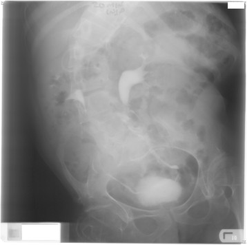
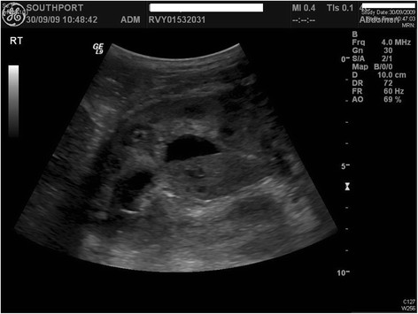
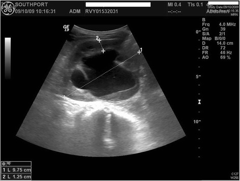
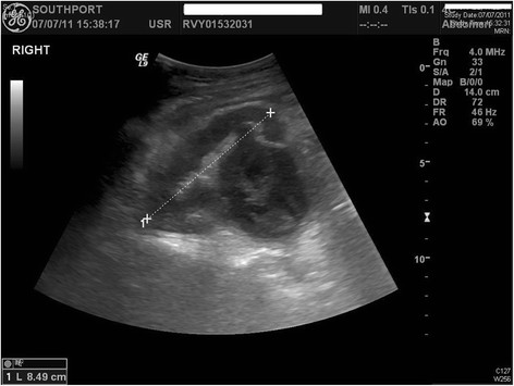
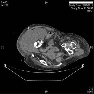
Case presentation: A 34-year-old male spinal bifida patient had been managing neuropathic bladder by penile sheath. Intravenous urography revealed normal kidneys. This patient was advised intermittent catheterisations. But self-catheterisation was not possible because of long, overhanging prepuce and marked spinal curvature. This patient developed repeated urine infections. Five years later, ultrasound examination of urinary tract revealed hydronephrotic right kidney with echogenic debris within the collecting system. There was no evidence of dilatation of the ureter near the vesicoureteric junction. The left kidney appeared normal. There was no evidence of calculus disease seen in either kidney. Indwelling urethral catheter drainage was established.Two years later, MAG-3 renogram revealed normal uptake and excretion by left kidney. The right kidney showed little functioning tissue. Following a routine change of urethral catheter this patient became unwell. Ultrasound examination revealed hydronephrotic right kidney containing thick hyper-echoic internal septations and debris in the right renal pelvis suspicious of pyonephrosis. Under both ultrasound and fluoroscopic guidance, an 8 French pig tail catheter was inserted into the right renal collecting system. 150 ml of turbid urine was aspirated immediately. This patient developed large left pleural effusion, collapse/consolidation of the left lower lobe, a large fluid collection in the abdomen extending into the pelvis and expired twenty days later because of sepsis and respiratory failure.
Conclusion: Although penile sheath drainage may be convenient for a spina bifida patient and the carers, hydronephrosis can occur insidiously. With recurrent urine infections, hydronephrotic kidney can become pyonephrosis, which is life-threatening. Therefore, every effort should be made to carry out intermittent catheterisations along with antimuscarinic drug therapy.
Figures
Similar articles
-
Long-term nephrostomy in an adult male spinal cord injury patient who had normal upper urinary tracts but developed bilateral hydronephrosis following penile sheath drainage: pyeloplasty and balloon dilatation of ureteropelvic junction proved futile: a case report.Cases J. 2009 Dec 16;2:9335. doi: 10.1186/1757-1626-2-9335. Cases J. 2009. PMID: 20062594 Free PMC article.
-
Recurrent bilateral renal calculi in a tetraplegic patient.Spinal Cord. 1998 Jul;36(7):454-62. doi: 10.1038/sj.sc.3100677. Spinal Cord. 1998. PMID: 9670380
-
Hydronephrosis and renal failure following inadequate management of neuropathic bladder in a patient with spinal cord injury: Case report of a preventable complication.Patient Saf Surg. 2012 Sep 26;6(1):22. doi: 10.1186/1754-9493-6-22. Patient Saf Surg. 2012. PMID: 23014062 Free PMC article.
-
Prevention of chronic kidney disease in spina bifida.Int Urol Nephrol. 2012 Jun;44(3):817-27. doi: 10.1007/s11255-010-9894-5. Epub 2011 Jan 13. Int Urol Nephrol. 2012. PMID: 21229390 Review.
-
Update on Urological Management of Spina Bifida from Prenatal Diagnosis to Adulthood.J Urol. 2015 Aug;194(2):288-96. doi: 10.1016/j.juro.2015.03.107. Epub 2015 Apr 1. J Urol. 2015. PMID: 25839383 Review.
Cited by
-
Inflammatory abdominal aortic aneurysm: a persistent painful hip.BMJ Case Rep. 2013 Sep 13;2013:bcr2013009150. doi: 10.1136/bcr-2013-009150. BMJ Case Rep. 2013. PMID: 24038286 Free PMC article.
-
Analysis of Nephrolithiasis Treatment in Highest Reference Hospital-Occurrence of Acromegaly in the Study Group.J Clin Med. 2023 Jun 6;12(12):3879. doi: 10.3390/jcm12123879. J Clin Med. 2023. PMID: 37373574 Free PMC article.
-
The 5th anniversary of "Patient Safety in Surgery" - from the Journal's origin to its future vision.Patient Saf Surg. 2012 Oct 18;6(1):24. doi: 10.1186/1754-9493-6-24. Patient Saf Surg. 2012. PMID: 23075037 Free PMC article. No abstract available.
References
-
- Parekh AD, Trusler LA, Pietsch JB, Byrne DW, DeMarco RT, Pope JC, Adams MC, Deshpande JK, Brock JW. Prospective, longitudinal evaluation of health related quality of life in the pediatric spina bifida population undergoing reconstructive urological surgery. J Urol. 2006;176(4 Pt 2):1878–1882. - PubMed
LinkOut - more resources
Full Text Sources
Research Materials
