. 2012 May 21:5:17.
doi: 10.1186/1756-6606-5-17.

Astrocytes generated from patient induced pluripotent stem cells recapitulate features of Huntington's disease patient cells

Affiliations

Astrocytes generated from patient induced pluripotent stem cells recapitulate features of Huntington's disease patient cells

Tarja A Juopperi et al. Mol Brain. .

Abstract

Background: Huntington's Disease (HD) is a devastating neurodegenerative disorder that clinically manifests as motor dysfunction, cognitive impairment and psychiatric symptoms. There is currently no cure for this progressive and fatal disorder. The causative mutation of this hereditary disease is a trinucleotide repeat expansion (CAG) in the Huntingtin gene that results in an expanded polyglutamine tract. Multiple mechanisms have been proposed to explain the preferential striatal and cortical degeneration that occurs with HD, including non-cell-autonomous contribution from astrocytes. Although numerous cell culture and animal models exist, there is a great need for experimental systems that can more accurately replicate the human disease. Human induced pluripotent stem cells (iPSCs) are a remarkable new tool to study neurological disorders because this cell type can be derived from patients as a renewable, genetically tractable source for unlimited cells that are difficult to acquire, such as neurons and astrocytes. The development of experimental systems based on iPSC technology could aid in the identification of molecular lesions and therapeutic treatments.

Results: We derived iPSCs from a father with adult onset HD and 50 CAG repeats (F-HD-iPSC) and his daughter with juvenile HD and 109 CAG repeats (D-HD-iPSC). These disease-specific iPSC lines were characterized by standard assays to assess the quality of iPSC lines and to demonstrate their pluripotency. HD-iPSCs were capable of producing phenotypically normal, functional neurons in vitro and were able to survive and differentiate into neurons in the adult mouse brain in vivo after transplantation. Surprisingly, when HD-iPSCs were directed to differentiate into an astrocytic lineage, we observed the presence of cytoplasmic, electron clear vacuoles in astrocytes from both F-HD-iPSCs and D-HD-iPSCs, which were significantly more pronounced in D-HD-astrocytes. Remarkably, the vacuolation in diseased astrocytes was observed under basal culture conditions without additional stressors and increased over time. Importantly, similar vacuolation phenotype has also been observed in peripheral blood lymphocytes from individuals with HD. Together, these data suggest that vacuolation may be a phenotype associated with HD.

Conclusions: We have generated a unique in vitro system to study HD pathogenesis using patient-specific iPSCs. The astrocytes derived from patient-specific iPSCs exhibit a vacuolation phenotype, a phenomenon previously documented in primary lymphocytes from HD patients. Our studies pave the way for future mechanistic investigations using human iPSCs to model HD and for high-throughput therapeutic screens.


Figures

Figure 1
Figure 1
Characterization of induced pluripotent stem cell lines (iPSCs). A, B: iPSC lines generated from fibroblasts exhibited typical ESC morphology by phase contrast microscopy (A) and maintained a normal karyotype (B). C: iPSCs strongly expressed the pluripotency marker alkaline phosphatase, whereas the surrounding MEFs were negative. D: Hematoxylin and eosin staining of teratomas derived from iPSC lines confirmed pluripotency by the presence of tissues representing the three major germ layers: ectoderm, endoderm, mesoderm. Scale bars: 100 μm.
Figure 2
Figure 2
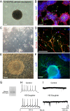
Differentiation of D-HD-iPSCs into neural progenitors, immature and mature functional neurons. A: A sample image of a neurosphere generated from the D-HD-iPSC line using the feeder-free differentiation protocol. B: Nestin-positive neural progenitors displaying rosette formation and immature (TUJ1+) neurons. C: Phase contrast image of neurons derived from D-HD-iPSCs. D: Mature (MAP2ab+) and doublecortin (DCX+) expressing neurons. E: Neural progenitor derived from D-HD-iPSCs using the PA6 cell line. Stromal cell monolayer observed in the background. F: Large networks of TUJ1+ neurons emanating from neural progenitors using the PA6 stromal cell line for induction. Scale bars: 100 μm. G: D-HD-iPSC derived neurons express both mutant and wild type HTT proteins by Western blot analysis. H: Action potentials recorded from neurons derived from D-HD-iPSCs and from C1-iPSCs. I: Miniature excitatory post-synaptic currents (mEPSCs) recorded from iPSC-derived neurons.
Figure 3
Figure 3
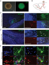
Transplantation and engraftment of neural progenitors derived from F-HD-iPSCs into the adult mouse brain. A: Neurospheres derived from F-HD-iPSCs expressing GFP prior to transplantation (phase contrast and fluorescence demonstrating GFP expression). B: A diagram illustrating neural progenitor cell transplantation. GFP-labeled neural progenitor cells were injected into the subventricular zone (SVZ) and migrated along the rostral migratory stream (RMS) to the olfactory bulb (OB). GFP and human nuclei antibody (HNA) positive neurons were detected in the RMS (C) at six weeks and in the OB (D) at eight weeks post-transplantation. E: NeuN+ GFP expressing cells exhibiting granule cell morphology were detected in the OB eight weeks post-transplantation. Unless otherwise indicated, scale bars: 100 μm.
Figure 4
Figure 4
Generation of astrocytes from iPSCs. A: A multi-step protocol was used to generate astrocytes. iPSCs were initially differentiated into neurospheres using a feeder-free protocol and subsequently differentiated into astrocytes. B: Astrocytes generated from HD-iPSCs exhibited a typical astrocyte morphology and expressed astrocyte markers glial fibrillary acidic protein (GFAP) and S100β. Scale bars: 100 μm (A) or 20 μm (B).
Figure 5
Figure 5
Astrocytes generated from the D-HD-iPSCs with increased cytoplasmic vacuolation. A: Vacuoles were detected in the cytoplasm of astrocytes (arrows) under phase contrast microscopy. The number of vacuoles varied depending on the iPSC line used to generate the astrocytes. B: Quantification of cytoplasmic vacuolation. Values represent mean + SEM (n = 4; *: P < 0.0001; ANOVA). C: Cytoplasmic vacuoles appeared empty and were negative for GFAP staining. D: The autophagosomal marker LC3 was weakly detected in astrocytes grown under normal culture conditions and rarely colocalized with cytoplasmic vacuoles. Vacuoles often appeared empty (arrows). Scale bars: 50 μm.
Figure 6
Figure 6
Enhanced vacuoles formation in astrocytes after chloroquine treatment. A: Overnight treatment of astrocytes with the autophagy inhibitor chloroquine resulted in increased cytoplasmic vacuolation for all cell lines examined, with D-HD-astrocytes exhibiting the largest number of vacuoles. B: S100β expressing astrocytes exhibited increased cytoplasmic staining with the autophagic vacuole marker LC3, after treatment with chloroquine. Scale bar: 50 μm.
Figure 7
Figure 7
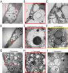
Ultrastructural changes in astrocytes derived from D-HD-iPSC. A: Numerous electron clear vacuoles were observed within the cytoplasm of astrocytes. B, C: A few autophagocytic vacuoles with myelin whorls (arrows) in astrocytes. D: Sample image of an astrocyte with many empty cytoplasmic vacuoles. Higher magnification revealed an autophagocytic vacuole with myelin whorls (E) and large spherical cytoplasmic granules representing lipid droplets (F). G, H, I: Chloroquine treated astrocytes exhibited numerous cytoplasmic vacuolation and proliferation of lysosomes. Scale bars: 2 μm (A-D, G, H) or 500 nm (E, F, I).

References

    1. Gusella JF, MacDonald ME. Huntington’s disease: seeing the pathogenic process through a genetic lens. Trends Biochem Sci. 2006;31(9):533–540. doi: 10.1016/j.tibs.2006.06.009. - DOI - PubMed
    1. Ross CA, Tabrizi SJ. Huntington’s disease: from molecular pathogenesis to clinical treatment. Lancet Neurol. 2011;10(1):83–98. doi: 10.1016/S1474-4422(10)70245-3. - DOI - PubMed
    1. A novel gene containing a trinucleotide repeat that is expanded and unstable on Huntington’s disease chromosomes. Cell. 1993;72(6):971–983. doi: 10.1016/0092-8674(93)90585-E. - DOI - PubMed
    1. DiFiglia M, Sapp E, Chase KO, Davies SW, Bates GP, Vonsattel JP, Aronin N. Aggregation of huntingtin in neuronal intranuclear inclusions and dystrophic neurites in brain. Science. 1997;277(5334):1990–1993. doi: 10.1126/science.277.5334.1990. - DOI - PubMed
    1. Imarisio S, Carmichael J, Korolchuk V, Chen CW, Saiki S, Rose C, Krishna G, Davies JE, Ttofi E, Underwood BR. et al.Huntington’s disease: from pathology and genetics to potential therapies. Biochem J. 2008;412(2):191–209. doi: 10.1042/BJ20071619. - DOI - PubMed

Publication types

MeSH terms