. 2012 Jul 6;287(28):23294-305.
doi: 10.1074/jbc.M111.313684. Epub 2012 May 21.

Pin1 protein regulates Smad protein signaling and pulmonary fibrosis

Affiliations

Pin1 protein regulates Smad protein signaling and pulmonary fibrosis

Zhong-Jian Shen et al. J Biol Chem. .

Abstract

Interstitial pulmonary fibrosis is caused by the excess production of extracellular matrix (ECM) by Fb in response to TGF-β1. Here, we show that the peptidyl-prolyl isomerase Pin1 modulates the production of many pro- and antifibrogenic cytokines and ECM. After acute, bleomycin injury, Pin1(-/-) mice showed reduced, pulmonary expression of collagens, tissue inhibitors of metalloproteinases, and fibrogenic cytokines but increased matrix metalloproteinases, compared with WT mice, despite similar levels of inflammation. In primary fibroblasts, Pin1 was required for TGF-β-induced phosphorylation, nuclear translocation, and transcriptional activity of Smad3. In Pin1(-/-) cells, inhibitory Smad6 was found in the cytoplasm rather than nucleus. Smad6 knockdown in Pin1(-/-) fibroblasts restored TGF-β-induced Smad3 activation, translocation, and target gene expression. Therefore, Pin1 is essential for normal Smad6 function and ECM production in response to injury or TGF-β and thus may be an attractive therapeutic target to prevent excess scarring in diverse lung diseases.


Figures

FIGURE 1.
FIGURE 1.
Pin1−/− mice are resistant to BLM-induced lung collagen deposition. A, representative lung sections stained with trichrome for collagen deposition (blue) on day 14 after PBS (−) or BLM (+) challenge. +/+, Pin1 wild type; −/−, knockout. Images are shown at ×10 magnification. B, ImageJ quantification of total collagen staining shown in A. C, left, representative airway stained with trichrome for collagen deposition (blue). Scale bar, 100 μm. Right, ImageJ quantification of total collagen staining. *, p < 0.05 by Student's t test in a two-tailed analysis. No difference was identified between +/+ and −/− mice in base-line collagen levels after PBS treatment. D, qPCR analysis of collagens I, III, and V in lungs of control and BLM-challenged mice on day 14. The mRNA levels in untreated wild-type mice were set as 100% throughout unless otherwise specified. E, immunoblot (left) of collagens in lung of control and BLM-challenged mice on day 14. Right, ImageJ quantification of the immunoblots shown on the left. *, p < 0.05 between BLM-challenged wild type and knockout. No significant differences were identified between +/+ and −/− mice in base-line collagen mRNA and protein levels or after PBS treatment. Data shown are representative of three independent experiments and are expressed as the mean ± S.D. (error bars) of eight animals.
FIGURE 2.
FIGURE 2.
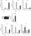
Pin1 regulates the expression of MMPs, TIMPs and fibrogenic growth factors in the lung. A, B, and D, qPCR analysis of ECM expression in the lung of control and BLM-challenged mice on day 14. **, p < 0.05 between vehicle-treated wild type and knockout. The FGF-1 mRNA data in D were all from BLM-treated mice (days 7 and 14). C, MMP2 (66 kDa) and MMP9 (84 kDa) bioactivity in BAL (n = 4–5) measured by zymography. NS, nonspecific bands. Right, ImageJ quantification of specific bands. *, p < 0.05 by Student's t test in a two-tailed analysis. Data shown are representative of three independent experiments and are expressed as the mean ± S.D. (error bars) of eight animals.
FIGURE 3.
FIGURE 3.
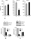
Pin1 knockout decreases type III collagen expression in primary lung Fb. A, wild-type and Pin1−/− primary Fb were starved for 2 days before stimulation with TGF-β1 (1 ng/ml) for 12 h. Total RNA were subjected to qPCR analysis. Data are shown as increased mRNA (percentage) after TGF-β1 compared with the respective untreated control. Error bars, S.D. of 3–4 separate cultures in each group. Data are representative of at least three independent experiments. *, p < 0.05 by Student's t test in a two-tailed analysis. B, cells were treated as in A, and secreted collagens in the culture medium were immunoblotted (top) with anti-collagen I and III. ImageJ quantification (bottom) of the immunoblots from two independent experiments. C, cell lysates were immunoblotted (top) with anti-collagen antibodies shown. ImageJ quantification (bottom) of the immunoblots from two independent experiments.
FIGURE 4.
FIGURE 4.
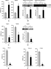
Pin1 regulates the expression of ECM and fibrogenic growth factors in primary lung Fb. Cells were treated as in Fig. 3A and analyzed for the expression of ECM and growth factors. A, mRNA for MMP2 and MMP3. Data are shown as increased mRNA (percentage) after TGF-β1 compared with respective untreated control. B, culture media from A were immunoblotted with anti-MMP-2, and the bioactivity was measured by zymography. C, mRNA for TIMP1 and TIMP4. Data are shown as increased mRNA (percentage) after TGF-β1 compared with respective untreated control. D, culture media from C were immunoblotted with anti-TIMP1. E and F, basal level of growth factor mRNA. Error bars, S.D. of 3–4 separate cultures in each group. Data are representative of at least five independent experiments *, p < 0.05 by Student's t test in a two-tailed analysis.
FIGURE 5.
FIGURE 5.
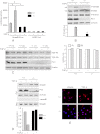
Pin knockout suppresses TGF-β1-Smad signaling in primary lung Fb. A, cells were infected with lentiviral vectors containing Smad2/3-responsive luciferase reporter and Renilla-luciferase constitutive reporter. After 2 days, cultures were treated with vehicle or TGF-β1 (1 ng/ml) for an additional 2 days. Cell lysates were subjected to luciferase activity assay using the Dual-Luciferase Assay system and normalized to the Renilla-luciferase reporter. Lenti-pGEX-luc without Smad2/3 binding sites served as negative control. B, cells were starved for 2 days and treated with TGF-β1 for 1 h. Cell lysates were immunoblotted with antibodies to phospho-Smad3 (Ser534/536), total Smad3, phospho-Erk1/2, Pin1, and β-actin. The ratios of Smad3 phosphorylation to the total Smad3 were quantified by ImageJ (bottom). AU, arbitrary units. C, cells from two mice were treated as in B, and total cell lysates were immunoblotted with anti-type I and II TGF-β1 receptors. ImageJ quantification of the immunoblots is shown on the right. D, cells were treated as in B, and cytoplasmic (Cyt) and nuclear (Nuc) extracts were immunoblotted (top) with antibodies to total Smad3, Pin1, β-actin, and lamin B1 (nuclear marker) (Nuc). C+, positive control. Bottom, ImageJ quantification of the immunoblots from two independent experiments. E, cells were treated as in B and immunostained with anti-Smad3 (red) and TO-Pro (nuclear dye; blue). *, p < 0.05. Data shown are representative of five independent experiments and are expressed as the mean ± S.D.
FIGURE 6.
FIGURE 6.
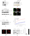
Pin1 directly interacts with Smad6 and modulates its localization. A, cell lysates were precleared with normal IgG and immunoprecipitated (IP) with anti-Pin1 prior to immunoblots with antibodies shown. 10% of lysates served as input. B, recombinant WT and mutant (MT) Smad6 with four potential Pin1 sites (Ser274-Pro, Ser283-Pro, Ser322-Pro, and Ser414-Pro) mutated from serine to glutamic acid were pulled down with GST and GST-Pin1 and probed with anti-Smad6. The inputs of control GST and GST-Pin1 were stained by Ponceau S (red) and are shown at the bottom. C, Pin1 interacts with the Ser274-Pro motif in Smad6. TAT-WT-Smad6 and TAT mutant Smad6 (S274A, S283A, and S322A) were added to the Fb culture before treatment of the cells with TGF-β1 (1 ng/ml) for 1 h. Cell lysates were pulled down with anti-Pin1 followed by immunoblot with anti-Pin1 and anti-His (Smad6). D, starved, wild-type cells (2 days) were treated with TGF-β1 for 1 h followed by immunostaining with anti-Pin1 (green), Smad6 (red), and TO-Pro (blue). E, cells were treated as in D and analyzed as in A. F, cells were treated with TGF-β1 for 10 min, and cytosolic protein was subjected to a Pin1 isomerase assay as described under “Experimental Procedures.” The products were measured at 390 nm. G, cells were treated as in D and immunostained with anti-Smad6. H, cells were treated as in D, and cytoplasmic (cyt) and nuclear extracts (nuc) were immunoblotted with antibody to Smad6, Pin1, and lamin B1 (nuclear marker). Data shown are representative of five independent experiments.
FIGURE 7.
FIGURE 7.
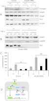
Smad6 knockdown restores TGF-β signaling in Pin1 null cells. A, primary lung Fb transfected with control or Smad6-specific siRNA (50 nm) were treated with TGF-β1 for 1 h. Whole cell lysates were immunoblotted with antibodies shown on the right. B, cells were treated as in A, and cytoplasmic (cyt) and nuclear extracts (nuc) were probed with antibodies shown on the right. Lamin B1 served as nuclear marker. C, cells were transfected with Smad6-specific siRNA and then infected with lentiviral vectors containing Smad2/3-responsive luciferase reporter and Renilla-luciferase constitutively active reporter. After 1 day, cultures were treated with vehicle or TGF-β1 (1 ng/ml) for an additional 2 days. Cell lysates were subjected to a luciferase activity assay using the Dual-Luciferase Assay system and normalized to the Renilla-luciferase reporter. Data shown are representative of at least five independent experiments and are expressed as the mean ± S.D. *, p < 0.05. NS, not significant. D, proposed model of Pin1 regulation of TGF-β/Smad signaling (for details, see “Results”).

Similar articles

Cited by

References

    1. Laurent G. J. (1987) Dynamic state of collagen: pathways of collagen degradation in vivo and their possible role in regulation of collagen mass. Am. J. Physiol. 252, C1–C9 - PubMed
    1. Wilson M. S., Madala S. K., Ramalingam T. R., Gochuico B. R., Rosas I. O., Cheever A. W., Wynn T. A. (2010) Bleomycin and IL-1β-mediated pulmonary fibrosis is IL-17A-dependent. J. Exp. Med. 207, 535–552 - PMC - PubMed
    1. Uhal B. D., Kim J. K., Li X., Molina-Molina M. (2007) Angiotensin-TGF-β1 cross-talk in human idiopathic pulmonary fibrosis. Autocrine mechanisms in myofibroblasts and macrophages. Curr. Pharm. Des. 13, 1247–1256 - PubMed
    1. Zheng L., Walters E. H., Wang N., Whitford H., Orsida B., Levvey B., Bailey M., Williams T. J., Snell G. I. (2004) Effect of inhaled fluticasone propionate on BAL TGF-β1 and bFGF concentrations in clinically stable lung transplant recipients. J. Heart Lung Transplant. 23, 446–455 - PubMed
    1. Halwani R., Al-Muhsen S., Al-Jahdali H., Hamid Q. (2011) Role of transforming growth factor-β in airway remodeling in asthma. Am. J. Respir. Cell Mol. Biol. 44, 127–133 - PubMed

Publication types

MeSH terms