. 2012 Jul 13;287(29):24082-91.
doi: 10.1074/jbc.M112.361485. Epub 2012 May 21.

The melanosomal protein PMEL17 as a target for antibody drug conjugate therapy in melanoma

Affiliations

The melanosomal protein PMEL17 as a target for antibody drug conjugate therapy in melanoma

Youjun Chen et al. J Biol Chem. .

Abstract

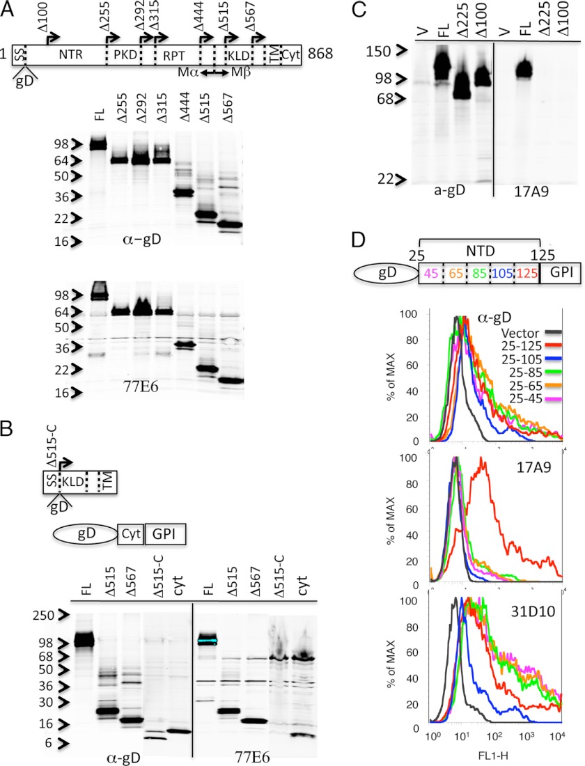
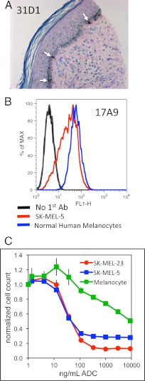
Melanocytes uniquely express specialized genes required for pigment formation, some of which are maintained following their transformation to melanoma. Here we exploit this property to selectively target melanoma with an antibody drug conjugate (ADC) specific to PMEL17, the product of the SILV pigment-forming gene. We describe new PMEL17 antibodies that detect the endogenous protein. These antibodies help define the secretory fate of PMEL17 and demonstrate its utility as an ADC target. Although newly synthesized PMEL17 is ultimately routed to the melanosome, we find substantial amounts accessible to our antibodies at the cell surface that undergo internalization and routing to a LAMP1-enriched, lysosome-related organelle. Accordingly, an ADC reactive with PMEL17 exhibits target-dependent tumor cell killing in vitro and in vivo.


Figures

FIGURE 1.
FIGURE 1.
Expression of PMEL17 mRNA. Measurements were carried out on the Affymetrix U133P chip and are expressed as scaled average difference. A, relative expression of PMEL17 in human tissues. Dots above and below the horizontal lines represent a specimen from the indicated normal or neoplastic tissue, respectively. B, relative expression of PMEL17 mRNA in cultured human cancer cell lines. BC, breast cancer; CRC, colorectal cancer; GB, glioblastoma; NSCLC, non-small cell lung cancer; SC, small cell lung cancer; NHL, Non-Hodgkin's lymphoma; MEL, melanoma; MM, multiple myeloma; OVCA, ovarian cancer; PANC, pancreatic cancer; PROS, prostate cancer. Only 5% of the 487 different cell lines analyzed are identified in the figure. For a complete list, see supplemental Table 1.
FIGURE 2.
FIGURE 2.
Binding of PMEL17 antibodies to intact cells. A, the melanoma cell lines SK-MEL-23, 1300mel, SK-MEL-5, and UACC257, normal melanocytes, and the prostate cancer cell line PC3 stably expressing PMEL17 were subjected to fluorescence-activated flow cytometry using antibody 17A9 (red line), 77E6 (blue line), and secondary antibody conjugated with phycoerythrin or the conjugated secondary antibody only (black line). B, fluorescence-activated cell sorting of mel928 cells stained with antibodies 17A9 and 77E6 directly labeled with Alexa Fluor 488 (vertical axis) and Alexa Fluor 555 (horizontal axis), respectively.
FIGURE 3.
FIGURE 3.
Localization of antibody binding sites on PMEL17. A, the PMEL17 deletion series represented in the schematic were expressed in mammalian cells, and the lysates were analyzed by immunoblotting with anti-gD and antibody 77E6. A signal sequence (SS) and the gD epitope were fused in-frame to each construct, followed by the PMEL17 sequence initiated at the indicated amino acid position (arrows). B, the Δ515 construct lacking the cytoplasmic domain (Δ515-C) and the cytoplasmic domain fused to a C-terminal GPI anchor (Cyt) were analyzed as in A. C, the indicated constructs were expressed and analyzed by immunoblotting with antibody 17A9. D, amino acid residues 25–125, 24–104, 25–85, 25–65, and 25–45 from the PMEL717 N-terminal domain (NTD) were fused to the gD epitope at the N terminus and GPI anchor at the C terminus. Constructs were expressed in 293 cells subjected to fluorescence-activated cell sorting with anti-gD (top panel), 17A9 (center panel), and 31D10 (bottom panel).
FIGURE 4.
FIGURE 4.
Immunfluorescent localization of PMEL17 in melanoma cells. A, fixed and permeabilized mel928 cells were reacted with antibodies 14C10 and 77E6 that were directly labeled with Alexa Fluor 488 (green) and Alexa Fluor 555 (red), respectively. Two examples are presented for the independent channels and the merged channel images. B, fluorescent activated cell sorting of mel928 cells stained with antibodies 17A9 and 77E6 directly labeled with Alexa Fluor 488 (light green) and Alexa Fluor 555 (red), respectively. Cells were also reacted with both antibodies simultaneously (blue), normal mouse IgG labeled with Alexa Fluor 488 (dark green), or Alexa Fluor 555 (fuchsia) or no antibody (black). Cytometry was gated for detection of Alexa Fluor 488 (left panel) or Alexa Fluor 555 (right panel). C, dual color confocal live imaging of mel928 cells reacted antibodies 17A9 and 77E6 that were directly labeled with Alexa Fluor 488 (green) and Alexa Fluor 555 (red), respectively.
FIGURE 5.
FIGURE 5.
Antibody internalization and cell killing. A, live 1300mel melanoma cells were incubated with mouse 17A9 for 2 h, fixed, permeabilized, and then reacted with rabbit-anti LAMP1 followed by anti-rabbit-Alexa Fluor 488 (LAMP1, green) and anti-mouse-Cy3 (17A9, red). B, the indicated cells were incubated with serial dilutions of the 17A9 ADC (ng/ml ADC), and 5 days later cell numbers were determined using CellTiter-GloII.
FIGURE 6.
FIGURE 6.
Expression of PMEL17 on normal melanocytes. A, PMEL17 expression in a fixed paraffin-embedded section of normal human skin was stained with mouse anti-PMEL17 antibody 31D10 and hematoxylin and eosin. B, normal human melanocytes and SK-MEL-5 melanoma cells were reacted with chimeric 17A9 or no primary antibody followed by Alexa Fluor 488-labeled anti-human IgG and analyzed by flow cytometry. C, SK-MEL-5 and SK-MEL-23 melanoma cells or normal human melanocytes were incubated with serial dilutions of the 17A9 ADC, and 5 days later cell numbers were determined using CellTiter-GloII.
FIGURE 7.
FIGURE 7.
Relative expression of PMEL17 on melanoma cells and ADC-dependent anti-tumor activity. A panel of 58 fixed paraffin-embedded specimens of human melanoma and five sections obtained from pelleted paraffin-embedded melanoma cell lines were stained with anti-PMEL17 antibodies 17A9 or 31D10. Scores for relative staining intensity (0,1, 2, or 3) for the melanomas are listed (table, bottom panel), and representative images for each scoring level are presented next to images of the five stained melanoma cell lines. B, subcutaneous tumors were established in mice inoculated with SK-MEL-23 cells. When tumor volumes reached ∼200 mm3 (day 0), animals were given a single intravenous injection of PBS (vehicle), control ADC (anti-GP120vcMMAE), or anti-PMEL17 ADC (17A9vcMMAE) at either 2 or 6 mg/kg (MPK). Average tumor volumes with standard deviations were determined from 10 animals per group.

References

    1. Garrett C. R., Eng C. (2011) Cetuximab in the treatment of patients with colorectal cancer. Expert Opin. Biol. Ther. 11, 937–949 - PubMed
    1. Alley S. C., Okeley N. M., Senter P. D. (2010) Antibody-drug conjugates. Targeted drug delivery for cancer. Curr. Opin. Chem. Biol. 14, 529–537 - PubMed
    1. Wilhite S. E., Barrett T. (2012) Strategies to explore functional genomics data sets in NCBI's GEO database. Methods Mol. Biol. 802, 41–53 - PMC - PubMed
    1. Schallreuter K. U., Kothari S., Chavan B., Spencer J. D. (2008) Regulation of melanogenesis. Controversies and new concepts. Exp. Dermatol. 17, 395–404 - PubMed
    1. Yamaguchi Y., Hearing V. J. (2009) Physiological factors that regulate skin pigmentation. Biofactors 35, 193–199 - PMC - PubMed

MeSH terms