. 2012 Aug 30:218:185-95.
doi: 10.1016/j.neuroscience.2012.05.022. Epub 2012 May 18.

Cholecystokinin knock-down in the basolateral amygdala has anxiolytic and antidepressant-like effects in mice

Affiliations

Cholecystokinin knock-down in the basolateral amygdala has anxiolytic and antidepressant-like effects in mice

C Del Boca et al. Neuroscience. .

Abstract

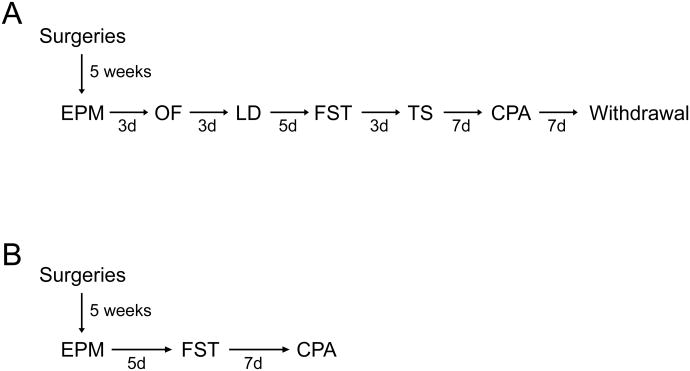
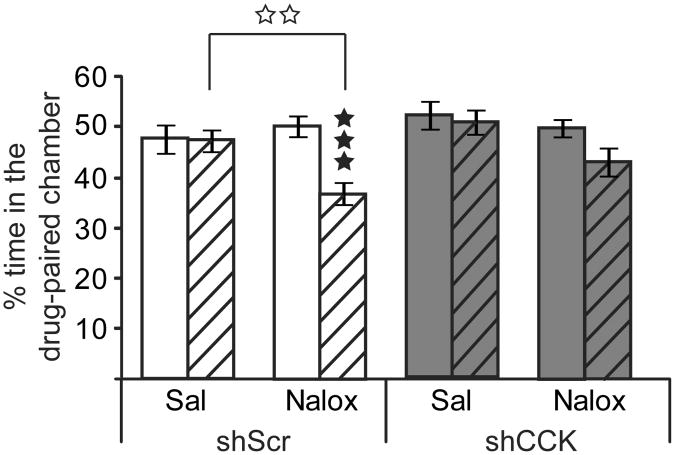
Cholecystokinin (CCK) is a neuropeptide widely distributed in the mammalian brain. This peptide regulates many physiological functions and behaviors, such as cardio-respiratory control, thermoregulation, nociception, feeding, memory processes and motivational responses, and plays a prominent role in emotional responses including anxiety and depression. CCK-expressing brain regions involved in these functions remain unclear and their identification represents an important step towards understanding CCK function in the brain. The basolateral amygdala (BLA) is strongly involved in emotional processing and expresses high levels of CCK. In this study we examined the contribution of CCK expressed in this brain region to emotional responses in mice. To knockdown CCK specifically in the BLA, we used stereotaxic delivery of recombinant adeno-associated viral vectors expressing a CCK-targeted shRNA. This procedure efficiently reduced CCK levels locally. shCCK-treated animals showed reduced levels of anxiety in the elevated plus-maze, and lower despair-like behavior in the forced swim test. Our data demonstrate that CCK expressed in the BLA represents a key brain substrate for anxiogenic and depressant effects of the peptide. The study also suggests that elevated amygdalar CCK could contribute to panic and major depressive disorders that have been associated with CCK dysfunction in humans.


Figures

Figure 1
Figure 1. Behavioral testing protocol to evaluate effects of CCK knock-down in the mouse BLA using two independent mouse cohorts
(A) Cohort 1. (B) Cohort 2 Abbreviations: EPM, Elevated Plus-Maze; OF, Open Field; LD, Light/Dark box; FST, Forced Swim Test; CPA, Naloxone induced-Conditioned Place Aversion; Naloxone precipitated-Withdrawal; d, days.
Figure 2
Figure 2. AAV2-shCCK injections in the BLA reduces CCK mRNA levels
Representative brain microsections showing DAPI staining (left), dig-labeled CCK mRNA (middle), and eGFP expression (right) of mice unilaterally injected into BLA with either AAV2-eGFP (top panels) or AAV2-shCCK (bottom panels) viral vectors (1.5μl, 3×10¹¹vg/ml). Mice were sacrificed for tissue collection either 2 or 6 weeks following surgery. CCK mRNA downregulation is observed in AAV2-shCCK but not AAV2-eGFP control samples, in an area overlapping maximal viral expression.
Figure 3
Figure 3. CCK knockdown in mouse BLA reduces levels of anxiety in the elevated plus-maze
Mice were bilaterally injected into BLA with either AAV2-shCCK (grey bars) or AAV2-shScramble (white bars) viral vectors and tested in the EPM. (A) Anxiety-like behavior. AAV2-shCCK-injected mice showed lower levels of anxiety, as indicated by time spent and number of entries in open (full bars) vs closed (striped bars) arms. Ratios of the time spent in and the number of entries to the open arms ((open arm)/(open arm + closed arms)) and total distance travelled are also shown. (B) Risk-taking behavior. AAV2-shCCK-injected mice showed increased risk-taking behavior as determined by number of total head dips in the open arms, number of head dips from the distal part of the open arms (last 1/3 of the arm) and percentage time spent in the distal part of the open arms. All results are expressed as mean ± SEM. Black stars, comparisons between arms; White stars, comparisons between shRNA. ★★★p<0.001, ✩p<0.05, ✩✩p<0.01, ✩✩✩p<0.001 (two-way ANOVA;n= 22 mice/group).
Figure 4
Figure 4. CCK knockdown in mouse BLA reduces despair-like behavior in the forced swim test
Mice were bilaterally injected into BLA with either AAV2-shCCK (grey bars and circles) or AAV2-shScramble (white bars and circles) viral vectors and tested in the FST. AAV2-shCCK-injected mice showed reduced despair-like behavior in both Cohort 1 (A) and Cohort 2 (B) as evaluated by percentage of immobility time during the 6 min test, time course of immobility in 2 min bins (seconds) and latency to the first immobility episode (seconds). For Cohort 2, data from repeated FST on day 1 and 2 are shown. All results are expressed as mean ± SEM. Black stars, individual comparisons between shRNA; asterisks, global time courses comparisons between shRNAs; white stars, comparisons between days. ★p<0.05, ★★p<0.01, ★★★p<0.001, **p<0.01, ***p<0.001, ✩✩p<0.01, ✩✩✩p<0.001 (one- and two-way ANOVA; n=10-11 mice/group in cohort 1, 9-10 mice/group in cohort 2).
Figure 5
Figure 5. CCK knockdown in mouse BLA reduces naloxone-induced place aversion (CPA)
Mice were bilaterally injected into BLA with either AAV2-shCCK (grey bars) or AAV2-shScramble (white bars) viral vectors. Naloxone place conditioning is shown as percentage of time spent in the drug-paired compartment during pre- (full bars) and post-conditioning (striped bars) sessions. Post-hoc analyses show that naloxone produced significant CPA in AAV2-shScramble but not in AAV2-shCCK mice. Results are expressed as mean ± SEM. Black stars, individual comparisons between sessions; white stars, individual comparisons between treatments. ★★★p<0.001; ✩✩p<0.01 (three-way ANOVA; n= 11 mice/group).
Figure 6
Figure 6. CCK knockdown in mouse BLA does not modify naloxone-precipitated withdrawal after chronic morphine treatment
Mice were bilaterally injected into BLA with either AAV2-shCCK (grey bars) or AAV2-shScramble (white bars) viral vectors, subjected to a chronic morphine regimen and naloxone-precipitated withdrawal. The global withdrawal score is shown ((horizontal activity + rearings + scratches + genital licks + head shakes + wet dog shakes + grooming) × 0.5 + (jumps + front paw tremors + sniffings) × 1 + (body tremors + ptosis + teeth chattering + piloerection) × 1), expressed as mean ± SEM. Naloxone-precipitated withdrawal did not differ between the two groups. ★★★p<0.01 (two-way ANOVA; n= 4 mice/group).
Figure 7
Figure 7. Quantitative validation of AAV2-shRNA-CCK knockdown into the mouse BLA
Mice were sacrificed for tissue collection 9 weeks after surgery. (A) Images from the Mouse Brain Atlas of Paxinos and Franklin (2001) showing histological reconstruction of injection sites into BLA. Black circles: correct injections of shCCK and shScramble vectors; red circles: .misplaced injections. (B) Representative brain sections showing eGFP expression (up), dig-labeled CCK mRNA (middle) and DAPI staining (bottom) from mice bilaterally injected into BLA with either AAV2-shScramble (left), or AAV2-shCCK (right) viral vectors (1.5μl, 3×10¹¹vg/ml). As in Figure 2, lower CCK mRNA is observed at the site of maximal viral expression in shCCK, but not shScramble, samples. (C) Left; representative autoradiographic image of a brain section processed for radioactive-ISH showing regions of interest (ROI) used in the quantification (Ctx, RAS cortex region; BG, Striatum BackGround; BLA, BasoLateral nucleus of the Amygdala). Right; quantification performed using a calibration curve of C14standards (ARC0146; American Radiolabeled Chemicals) and theImageJ software. A rodbard non-lineal regression function was used to convert grey levelsinto counts of radioactivity (nCi/mg). CCK mRNA expression is significantly reduced in theBLA of AAV2-shCCK treated animals (grey bar) compared to their AAV2-shScramble controls (white bar). Results are expressed in normalized values obtained from meanradioactive measures [(BLA − Striatum background)/(RAS Cortex − Striatum background)] ±SEM. ★★p<0.01 (Unpaired Student's t-test; n=5 mice/condition).

References

    1. Ambesi-Impiombato A, D'Urso G, Muscettola G, de Bartolomeis A. Method for quantitative in situ hybridization histochemistry and image analysis applied for Homer1a gene expression in rat brain. Brain Res Brain Res Protoc. 2003;11:189–196. - PubMed
    1. Becker C, Zeau B, Rivat C, Blugeot A, Hamon M, Benoliel JJ. Repeated social defeat-induced depression-like behavioral and biological alterations in rats: involvement of cholecystokinin. Mol Psychiatry. 2008;13:1079–1092. - PubMed
    1. Beinfeld MC. An introduction to neuronal cholecystokinin. Peptides. 2001;22:1197–1200. - PubMed
    1. Belcheva I, Belcheva S, Petkov VV, Petkov VD. Asymmetry in behavioral responses to cholecystokinin microinjected into rat nucleus accumbens and amygdala. Neuropharmacology. 1994;33:995–1002. - PubMed
    1. Berna MJ, Tapia JA, Sancho V, Jensen RT. Progress in developing cholecystokinin(CCK)/gastrin receptor ligands that have therapeutic potential. Curr Opin Pharmacol. 2007;7:583–592. - PMC - PubMed

Publication types

Substances